ABSTRACT
Background
Medicinal plants have historically played a crucial role in healthcare, serving as the basis for many modern drugs. Among these, Drymaria cordata (L.) Willd. ex Schult., a small herbaceous plant of the Caryophyllaceae family, has garnered attention for its medicinal properties. This study investigates the hepatoprotective potential of its ethanolic extract and ethyl acetate fraction of Drymaria cordata (L.) Willd. ex Schult. against lipopolysaccharide-induced cytotoxicity in HepG2 cells.
Materials and Methods
The in vitro hepatoprotective properties of ethanolic extract and ethyl acetate fraction of Drymaria cordata (L.) Willd. ex Schult. were evaluated by measuring cell viability; activities of Superoxide dismutase, reduced glutathione, Catalase and Malondialdehyde, aspartate aminotransferase, alanine aminotransferase, and Alkaline phosphatase levels.
Results
The results demonstrated that both ethanolic extract and ethyl acetate fraction significantly restored Superoxide dismutase, reduced glutathione, Catalase and Malondialdehyde, aspartate aminotransferase, alanine aminotransferase, Alkaline phosphatase levels, while also preserving cell viability. Notably, the ethyl acetate fraction exhibited a stronger effect in lowering oxidative stress parameters, indicating its superior role in cellular antioxidant defense. Additionally, the study evaluated the phosphorylation status of Protein Kinase C through Sandwich ELISA, revealing a potential regulatory mechanism.
Conclusion
The findings highlight that the ethyl acetate fraction of Drymaria cordata (L.) Willd. ex Schult. may serve as a promising natural hepatoprotective, antioxidant agent alternative for managing conditions associated with Protein Kinase C activation and inflammation. Its potent antioxidant and hepatoprotective properties suggest that Drymaria cordata (L.) Willd. ex Schult. could be developed as a cost-effective therapeutic option.
INTRODUCTION
Liver diseases remain a major global health challenge, significantly contributing to morbidity and mortality worldwide (Schuppanet al., 2008). Hepatotoxicity, often resulting from oxidative stress, alcohol consumption, and infections, is a major pathological condition that can lead to severe complications (Thakuret al., 2024; Ferdeket al., 2022). Despite advancements in pharmacological treatments, the efficacy of current therapies for liver diseases remains limited, often accompanied by adverse side effects and high costs (Teschkeet al., 2015).
Medicinal plants have historically played a crucial role in healthcare, serving as the basis for many modern drugs (Newmanet al., 2020). Among these, Drymaria cordata (L.) Willd. ex Schult., a small herbaceous plant of the Caryophyllaceae family, has garnered attention for its medicinal properties. Traditionally used in ethnomedicine across tropical and subtropical regions, the plant has been employed to treat inflammatory disorders, wounds, and microbial infections (Singlaet al., 2023). Phytochemical analysis has revealed the presence of bioactive compounds like flavonoids, alkaloids, tannins, etc, which exhibit antioxidant, anti-inflammatory, and cytoprotective activities (Maran et al., 2022; Baruaet al., 2011). These characteristics suggest that Drymaria cordata (L.) Willd. ex Schult. could serve as a promising hepatoprotective agent. However, its hepatoprotective efficacy, particularly in the context of in vitro studies, remains underexplored. This study aims to investigate the in vitro hepatoprotective activity of Drymaria cordata (L.) Willd. ex Schult. using the HepG2 cell line. By analyzing parameters such as cell viability, oxidative stress markers, and enzymatic activity, the research seeks to provide scientific evidence supporting the traditional use of Drymaria cordata (L.) Willd. ex Schult. in liver-related ailments.
MATERIALS AND METHODS
Extraction and Fractionation
Fresh aerial parts of Drymaria cordata (L.) Willd. ex Schult. were collected in March 2023 from Mahmora, Charaideo, Assam, India. The specimen was authenticated at the Botany Department, Guwahati University (Accession No. GUBH20312). Dried aerial parts were extracted with 95% ethanol using a Soxhlet apparatus. The crude extract was fractionated by liquid-liquid extraction with petroleum ether and ethyl acetate. The concentrated extracts were dried under vacuum at 40-50ºC using a rotary evaporator and subsequently stored in a refrigerator for further analysis (Awadet al., 2015).
Cell Viability Assay (MTT Assay)
HepG2 cells (NCCS Pune) were cultured in DMEM with 10% FBS and 1% penicillin-streptomycin (100 μg/mL) under 5% CO₂. Cells were seeded in 96-well plates (4 × 10⁵ cells/well) and treated with varying concentrations (10-400 μg/mL) of EDC and EADC and 0.1% DMSO (control) for 24 hr. Afterward, 20 μl of 2.5 mg/mL MTT (3-[4,5-dimethylthiazol-2-yl]-2,5-diphenyl tetrazolium bromide) was added, followed by 4 hr of incubation. Formazan crystals were dissolved in 500 μL DMSO, and absorbance was measured at 570 nm to calculate cell viability (Zhanget al., 2019).
Measurement of superoxide dismutase activity
Estimation of Reduced glutathione level
The GSH level of cells were determined as per the reported method and expressed as ng/mg protein of tissue (Ellman, 1959). The total protein of cells was estimated as per the standard method (Lowryet al., 1951).
Estimation of Catalase Activity
CAT activities in HepG2 cells were assayed as per the reported methods (Aebi 1974). The CAT activity was expressed in terms of hydrogen peroxide decomposed in kU/min/mg protein tissue (Aebi, 1974).
Malondialdehyde Measurement
AST, ALT and ALP Activity
Colorimetric methods were employed to assess AST, ALT, and ALP activities, as elevated levels of these enzymes typically indicate liver tissue damage. HepG2 cells were seeded in 96-well plates at 2 × 10⁶ cells/well and incubated at 37ºC with 5% CO₂ until confluent. Cells were then maintained in DMEM with 2% FBS for 24 hr. Subsequently, cells were treated with or without 25 μg/mL LPS, Silymarin, EDC, and EADC (0-100 μg/mL) for 24 hr. AST, ALT, and ALP activities were measured using respective Elabscience kits (E-BC-K236-M, E-BC-K235-M, E-BC-K091-M) following manufacturer protocols. Results are expressed in IU/L (Pareeket al., 2013; Gonzálezet al., 2017).
Lipopolysaccharide-induced Phospho-PKC Activity in HepG2 Cells
EADC fraction of Drymaria cordata (L.) Willd. ex Schult. have been chosen for pPKC determination due the better antioxidant and hepatoprotective efficacy as compared to EDC. HepG2 cells (NCCS Pune) were cultured in DMEM supplemented with 10% FBS in a 5% CO₂ atmosphere under controlled conditions. Over 95% of viable cells were treated with or without LPS (25 μg/mL) and different concentrations (0-100 μg/mL) of EADC in 96-well plates. Phospho-PKC levels were assessed using a commercially available Sandwich ELISA kit (KBH15203) (Mauryaet al., 2015).
Statistical Analysis
The results of antioxidant and hepatoprotective activity were expressed as the mean ±SEM. A statistical analysis was performed with one-way analysis of variance followed by Tukey’s multiple comparison tests using GraphPad Prism 8.0.2 (263) 2019 Software. Differences were considered statistically significant at the value of probability less than 5% (p<0.05)
RESULTS
Cell Viability Assay (MTT Assay)
The percentage cell viability with respect to the normal control cell lines (HepG2) at different concentrations of EDC extract and EADC were determined. The normal control cells showed 100% cell viability in HepG2 cells. The EDC at concentrations 10 μg/mL, 25 μg/mL, 50 μg/mL, 100 μg/mL, 200 μg/mL and 400 μg/mL showed 95.24±2.61%, 92.44±4.01%, 87.44±4.28%, 80.10±2.70%, 78.53±3.42% and 77.65±2.44% cell viability, respectively. The EADC at concentrations 10 μg/mL, 25 μg/mL, 50 μg/mL, 100 μg/mL, 200 μg/mL and 400 showed 96.42±2.20%, 93.37±3.77%, 91.08±4.47%, 87.56±2.21%, 83.04±3.05% and 78.26±2.28% cell viability, respectively.
Measurement of Superoxide dismutase activity
Figures 1A and B represents the status of SOD activities in HepG2 cell lines with and without treatment of 25 μg/mL LPS and activities were 66.98±1.77 mU/min/mg protein and 125.78±1.16 mU/min/mg protein respectively. SOD activity was significantly increased in HepG2 cell line when different concentration of Silymarin, EDC and EADC (10, 25, 50, 75 and 100 µg/mL) were introduced in the cultures respectively.

Figure 1:
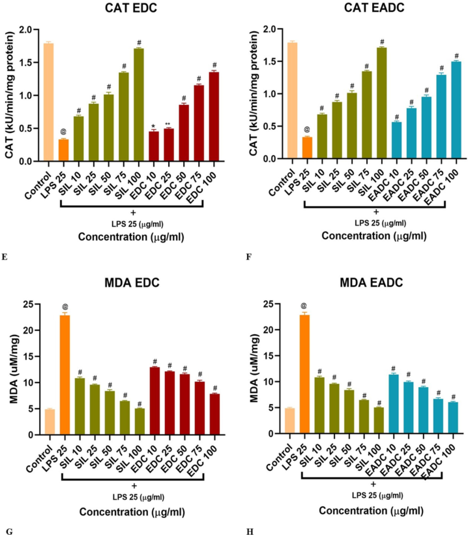
Antioxidant enzyme activities in Hepatoma cell line (HepG2). (A, B) SOD activity with and without treatment of EDC and EADC, respectively. (C, D) GSH activity with and without EDC and EADC treatment, respectively. (E, F) CAT activity with and without EDC and EADC treatment, respectively. (G, H) MDA levels with and without EDC and EADC treatment, respectively. Data are expressed as mean±SEM (n=3). @p<0.0001 compared with control group; #p<0.0001, ***p<0.001, **p<0.01, *p<0.05 compared with LPS-treated group.
Estimation of Reduced glutathione level
Figures 1C and D depict the GSH content in HepG2 cell lines with and without treatment of 25 μg/mL LPS, and levels were 184.01±8.04 ng/mg protein and 577.94±13.04 ng/mg protein, respectively. GSH levels were significantly increased in HepG2 cell lines when different concentrations of Silymarin, EDC, and EADC (10, 25, 50, 75, and 100 µg/mL) were introduced into the cultures, respectively.
Estimation of Catalase Activity
Figures 1E and F represent the status of CAT activities in HepG2 cell lines with and without treatment of 25 μg/mL LPS, and levels were 0.33±0.01 kU/min/mg protein and 1.79±0.03 kU/min/mg protein, respectively. CAT activities were significantly increased in HepG2 cell lines when different concentrations of Silymarin, EDC, and EADC (10, 25, 50, 75, and 100 µg/mL) were introduced into the cultures, respectively.
Malondialdehyde Measurement
Figures 1G and H depict the status of MDA in HepG2 cell lines with and without treatment of 25 μg/mL LPS, and levels were 23.87±0.19 uM/mg and 4.89±0.17 uM/mg, respectively. MDA levels were significantly reduced in HepG2 cell lines when different concentrations of Silymarin, EDC, and EADC (10, 25, 50, 75, and 100 µg/mL) were introduced in the cultures, respectively.
AST, ALT and ALP Activity
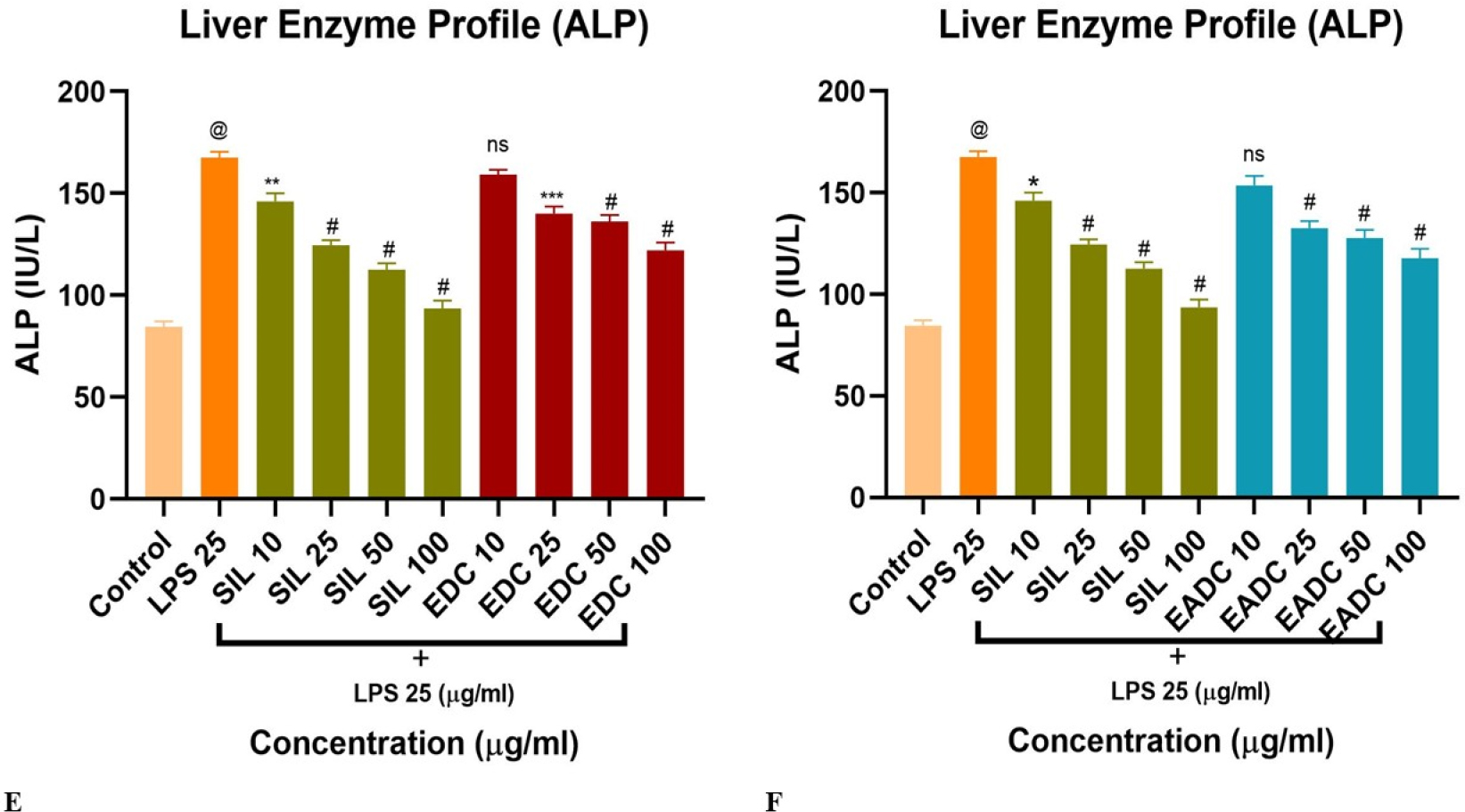
Figures 2A-F represent the level of AST, ALT and ALP respectively. A significant increase in the levels of AST, ALT and ALP as compared to control was observed in 25 µg/mL LPS exposed HepG2 cells. These cells, when treated with different concentrations of Silymarin, EDC, and EADC (10, 25, 50, and 100 µg/mL), showed a significant restoration in altered biochemical parameters towards the normal and were dose dependent.

Figure 2:
Effect of Drymaria cordata (L.) Willd. ex Schult. extracts on liver marker enzymes in LPS-induced HepG2 cells. (A, B) Effect of Ethanolic Extract (EDC) and Ethyl Acetate fraction (EADC) on AST activity, respectively. (C, D) Effect of EDC and EADC on ALT activity,respectively. (E, F) Effect of EDC and EADC on ALP activity, respectively. Data are expressed as mean±SEM (n=3). @p<0.0001 compared with control group; #p<0.0001, ***p<0.001, **p<0.01, *p<0.05 compared with LPS-treated group.
Lipopolysaccharide-induced Phospho-PKC Activity in HepG2 Cells
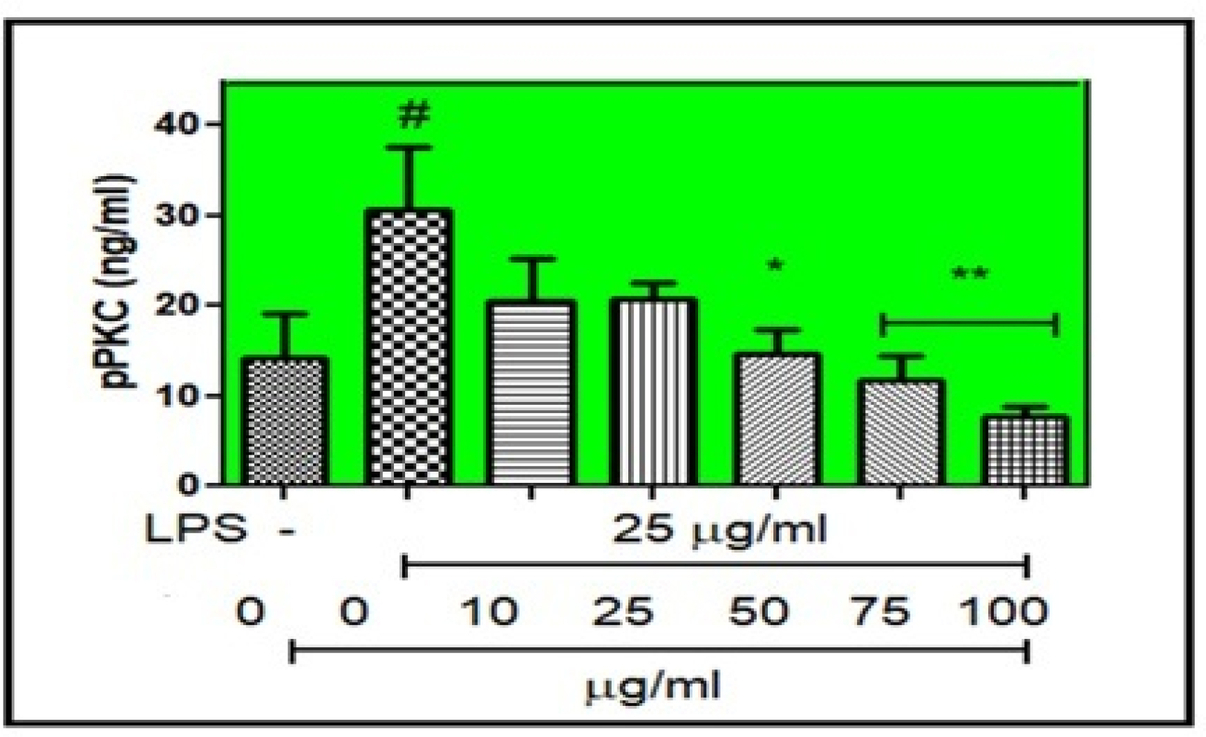
As the EADC fraction was more effective than the EDC extract on the liver function enzymes and oxidative stress markers, it was further used for the determination of phospho-PKC activity in HepG2 cells. When the pPKC of EADC was estimated by Sandwich ELISA, EADC exhibited a significant and profound outcome by the inhibition of phosphorylation of PKC. Figure 3 depicts the status of pPKC level in HepG2 cells with or without treatment of 25 μg/mL LPS, and levels were 30.54±6.98 ng/mL and 14.13±4.94 ng/mL, respectively.

Figure 3:
Effect of EADC on pPKC by Sandwich ELISA. Data were expressed as mean±SEM (n=4). #p<0.05 when compared with control (without LPS); *p<0.05, **p<0.01, when compared with control (with LPS).
DISCUSSION
The present study highlights the hepatoprotective effects of Drymaria cordata (L.) Willd. ex Schult. extracts, specifically the EDC and EADC, against LPS-induced toxicity in HepG2 cells. The MTT assay demonstrated that both EDC and EADC supported high cell viability, indicating their biocompatibility and potential protective effects. At lower concentrations (10-50 µg/mL), both fractions-maintained cell viability above 75%, suggesting minimal cytotoxicity. Even at moderate concentrations (100-200 µg/mL), cell survival remained high, reinforcing the protective potential of the extracts. However, at higher concentrations (400 µg/mL), a slight reduction in cell viability was observed, particularly in the EDC extract as compared to the EADC fraction, indicating a dose-dependent effect. The EADC fraction exhibited a more stable viability profile, suggesting its potential suitability for applications requiring minimal cytotoxic effects.
Oxidative stress plays a major role in hepatic injury, in order to assess Drymaria cordata (L.) Willd. ex Schult. extracts on oxidative stress markers such as: SOD, GSH, CAT and MDA. Following LPS exposure, SOD and GSH levels were markedly restored, identifying oxidative stress-induced cellular damage. Controlled treatment with EDC and EADC did indeed significantly recover those levels, especially at 50 and 100 µg/mL, suggesting that they induced a potent antioxidant response.
Antioxidant capability of the EADC fraction was marginally superior to that of the DC extract, indicating the existence of strong free radical-scavenging substances. Lipid peroxidation, evaluated through MDA, was notably increased in cells treated with LPS, indicating cellular stress and damage to membranes. Both EDC and EADC significantly lowered MDA levels, with the EADC fraction showing a stronger effect, suggesting its involvement in mitigating lipid peroxidation and oxidative damage. Reduced CAT activity following LPS exposure also indicated compromised antioxidant defense mechanisms. However, treatment of EDC and EADC restored CAT activity, particularly at concentrations of 50 and 100 µg/mL, highlighting their role in enhancing cellular antioxidant protection and protecting hepatocytes from oxidative damage.
Liver function enzyme analysis indicated that LPS exposure significantly elevated AST, ALT, and ALP levels, marking hepatic stress and injury. However, co-treatment with Drymaria cordata (L.) Willd. ex Schult. (EDC and EADC) extract and fraction effectively reduced these enzyme levels, suggesting notable hepatoprotective activity. The most effective concentrations (50 and 100 µg/mL) exhibited liver-protective properties comparable to the standard drug, silymarin. These findings suggest that Drymaria cordata (L.) Willd. ex Schult. extracts may contribute to restoring liver function and preserving hepatocyte integrity.
The EDC and EADC fractions showed considerable hepatoprotective and antioxidant properties, as indicated by a comparative assessment of the extracts; nonetheless, the EADC fraction was more successful in restoring oxidative stress markers such as SOD, GSH, MDA, and CAT, while also preserving cell viability. Improving focus might enhance treatment efficacy and lower possible risks, according to the observed dose-dependent effects.
Moreover, the phosphorylation status of Protein Kinase C (pPKC) was assessed via a Sandwich ELISA to investigate how Drymaria cordata (L.) Willd. ex Schult. fraction influence on PKC signalling in HepG2 cells. The findings indicated a significant impact of the EADC fraction on inhibiting PKC phosphorylation. In the control group, the baseline pPKC level was recorded at 14.13±4.94 ng/mL, whereas LPS treatment significantly increased pPKC levels to 30.54±6.98 ng/mL. This increase aligns with the established role of LPS in activating PKC signalling pathways, essential for inflammatory reactions and cellular stress mechanisms. Nonetheless, therapy with the EADC fraction significantly decreased pPKC levels, indicating its anti-inflammatory and cytoprotective effects. The observed decrease in pPKC following treatment indicates that the EADC fraction effectively hinders PKC phosphorylation, potentially disrupting inflammatory pathways triggered by LPS.
This result aligns with previous studies highlighting the important function of PKC in inflammation, as inhibiting its phosphorylation has been shown to decrease inflammation. These findings suggest that the EADC part of Drymaria cordata (L.) Willd. ex Schult. may serve as a possible treatment for conditions associated with increased PKC activation and inflammation. Further extensive studies, including mechanistic evaluations and in vivo validations, will be essential to completely assess its therapeutic potential.
CONCLUSION
Our findings confirm the hepatoprotective and antioxidant effects of Drymaria cordata (L.) Willd. ex Schult. extracts, particularly the ethyl acetate fraction, against LPS-induced cytotoxicity in HepG2 cells. The fractions demonstrated high cell viability at moderate concentrations, indicating significant cytoprotective potential. The ethyl acetate fraction also reduced pPKC levels, highlighting its anti-inflammatory properties, which are crucial in mitigating liver damage caused by inflammation. In conclusion, Drymaria cordata (L.) Willd. ex Schult., especially its ethyl acetate fraction, appears to be a promising candidate for hepatoprotection and antioxidant therapy. These findings support its potential application as a natural treatment for liver conditions associated with inflammation and oxidative stress.
Cite this article:
Yakin J, Alam F. In vitro Hepatoprotective Effect of Drymaria cordata Against Lipopolysaccharide-Induced Hepatotoxicity in HepG2 Cell Line. J Young Pharm. 2025;17(3):543-50.
ACKNOWLEDGEMENT
The authors are thankful to Assam down town University, for providing laboratory and the basic facilities for carrying out experimental work.
ABBREVIATIONS
| EDC | Ethanolic Drymaria cordata |
|---|---|
| EADC | Ethayl acetate Drymaria cordata |
| LPS | Lipopolysaccharide |
| AST | Aspartate Aminotransferase |
| ALT | Alanine Aminotransferase |
| ALP | Alkaline Phosphatase |
| SOD | Superoxide Dismutase |
| GSH | Reduced Glutathione |
| MDA | Malondialdehyde |
| CAT | Catalase |
| PKC | Protein Kinase C |
| pPKC | Phosphorylation of Protein Kinase C |
| ELISA | Enzyme-Linked Immunosorbent Assay |
| DMEM | Dulbecco’s Modified Eagle Medium |
| FBS | Fetal bovine serum |
| DMSO | Dimethyl Sulfoxide |
| SEM | Standard Error of the Mean. |
References
- Aebi H., Packer L.. (1974) Methods of enzymatic analysis 2: 673-684 https://doi.org/10.1016/B978-0-12-091302-2.50032-3
- Awad N. E., Abdelkawy M. A., Hamed M. A., Souleman A. M. A., Abdelrahman E. H., Ramadan N. S., et al. (2015) Antioxidant and hepatoprotective effects of ethyl acetate fraction and characterization of its anthocyanin content. International Journal of Pharmacy and Pharmaceutical Sciences 7: 91-96 https://doi.org/10.1016/B978-0-12-091302-2.50032-3 | Google Scholar
- Bala A., Haldar P. K., Kar B., Naskar S., Mazumder U. K.. (2012) Carbon tetrachloride: A hepatotoxin causes oxidative stress in murine peritoneal macrophage and peripheral blood lymphocyte cells. Immunopharmacology and Immunotoxicology 34: 157-162 https://doi.org/10.3109/08923973.2011.590498 | Google Scholar
- Barua C. C., Roy J. D., Buragohain B., Barua A. G., Borah P., Lahkar M., et al. (2011) Analgesic and anti-nociceptive activity of hydroethanolic extract of Willd. Indian Journal of Pharmacology 43: 121-125 https://doi.org/10.4103/0253-7613.77337 | Google Scholar
- Ellman G. L.. (1959) Tissue sulfhydryl groups. Archives of Biochemistry and Biophysics 82: 70-77 https://doi.org/10.1016/0003-9861(59)90090-6 | Google Scholar
- Ferdek P. E., Krzysztofik D., Stopa K. B., Kusiak A. A., Paw M., Wnuk D., Jakubowska M. A., et al. (2022) When healing turns into killing-The pathophysiology of pancreatic and hepatic fibrosis. The Journal of Physiology 600: 2579-2612 https://doi.org/10.1113/JP281135 | Google Scholar
- González L. T., Minsky N. W., Espinosa L. E. M., Aranda R. S., Meseguer J. P., Pérez P. C., et al. (2017)
In vitro assessment of hepatoprotective agents against damage induced by acetaminophen and CCl4. BMC Complementary and Alternative Medicine 17: 39 https://doi.org/10.1186/s12906-016-1506-1 | Google Scholar - Haldar P. K., Bhattacharya S., Bala A., Kar B., Mazumder U. K., Dewanjee S., et al. (2010) Chemopreventive role of against 20-methylcholanthrene-induced carcinogenesis in mouse. Toxicological and Environmental Chemistry 92: 1749-1763 https://doi.org/10.1080/02772241003783703 | Google Scholar
- Kakkar P., Das B., Viswanathan P. N.. (1984) A modified spectrophotometric assay of superoxide dismutase. Indian Journal of Biochemistry and Biophysics 21: 130-132 https://doi.org/10.1080/02772241003783703 | Google Scholar
- Lowry O. H., Rosebrough N. J., Farr A. L., Randall R. J.. (1951) Protein measurement with the Folin phenol reagent. Journal of Biological Chemistry 193: 265-275 https://doi.org/10.1016/S0021-9258(19)52451-6 | Google Scholar
- Maurya A. K., Vinayak M.. (2015) Anticarcinogenic action of quercetin by downregulation of phosphatidylinositol 3-kinase (PI3K) and protein kinase C (PKC) via induction of p53 in hepatocellular Carcinoma (HepG2) cell line. Molecular Biology Reports 42: 1419-1429 https://doi.org/10.1007/s11033-015-3921-7 | Google Scholar
- Newman D. J., Cragg G. M.. (2020) Natural products as sources of new drugs over the nearly four decades from 1981 to 2019. Journal of Natural Products 83: 770-803 https://doi.org/10.1021/acs.jnatprod.9b01285 | Google Scholar
- Ohkawa H., Ohishi N., Yagi K.. (1979) Assay for lipid peroxides in animal tissues by thiobarbituric acid reaction. Analytical Biochemistry 95: 351-358 https://doi.org/10.1016/0003-2697(79)90738-3 | Google Scholar
- Pareek A., Godavarthi A., Nagori B. P.. (2013)
In vitro hepatoprotective activity of Corchorus depressus L. against CCl₄-induced toxicity in HepG2 cell line. Pharmacognosy Journal 5: 191-195 https://doi.org/10.1016/j.phcgj.2013.07.001 | Google Scholar - Schuppan D., Afdhal N. H.. (2008) Liver cirrhosis. The Lancet 371: 838-851 https://doi.org/10.1016/S0140-6736(08)60383-9 | Google Scholar
- Singla S., Pradhan J., Thakur R., Goyal S.. (2023)
Drymaria cordata: Review on its pharmacognosy, phytochemistry, and pharmacological profile. Phytomedicine Plus 3: 1-12 https://doi.org/10.1016/j.phyplu.2023.100469 | Google Scholar - Teschke R., Eickhoff A.. (2015) Herbal hepatotoxicity in traditional and modern medicine: Actual key issues and new encouraging steps. Frontiers in Pharmacology 6: 72 https://doi.org/10.3389/fphar.2015.00072 | Google Scholar
- Thakur S., Kumar V., Das R., Sharma V., Mehta D. K.. (2024) Biomarkers of hepatic toxicity: An overview. Current Therapeutic Research, Clinical and Experimental 100: Article 100737 https://doi.org/10.1016/j.curtheres.2024.100737 | Google Scholar
- Venmathi Maran B. A., Iqbal M., Gangadaran P., Ahn B.-C., Rao P. V., Shah M. D., et al. (2022) Hepatoprotective potential of Malaysian medicinal plants: A review on phytochemicals, oxidative stress, and antioxidant mechanisms. Molecules 27: 1533 https://doi.org/10.3390/molecules27051533 | Google Scholar
- Zhang Q., Bao J., Yang J.. (2019) Genistein-triggered anticancer activity against liver cancer cell line HepG2 involves ROS generation, mitochondrial apoptosis, G2/M cell cycle arrest, and inhibition of cell migration. Archives of Medical Science 15: 1001-1009 https://doi.org/10.5114/aoms.2018.78742 | Google Scholar
