ABSTRACT
Background
Dental caries are highly prevalent and if left untreated, it can lead to harmful consequences. Micro-invasive types of dental caries treatment were adopted to cease the progression of decay. Fissure sealant helps to arrest incipient caries in pits and fissures. However, the major concern with conventional pit and fissure sealants is their technique sensitivity due to moisture contamination. Hence, hydrophilic sealants were introduced to overcome this drawback. The objective of the study is to compare and evaluate the micro hardness of conventional and hydrophilic pit and fissure sealants.
Materials and Methods
Thirty sound molar teeth were grouped into 2 groups for which 15 molars were assigned to each group. Group I was allocated for 3M ESPE Clinpro hydrophobic sealants and Group II was Ultra-seal XT Hydro hydrophilic sealant. The prepared specimens were acid-etched with 37% phosphoric acid, followed by rinsing with water and finally air-dried before sealant placement. Each sealant material was then applied and light-cured. Vickers hardness test was used to estimate the microhardness of the sample with 200 gm load for 20 sec. Mann Whitney U test was done to find the difference between the two groups and the Wilcoxon test was used to find the difference within the group.
Results
The differences between the mean microhardness value and Immediate, Aging time factor were found to be statistically non-significant. An increase in mean value was observed after ageing in both groups. A statistically significant difference (p <0.05) was observed within each group for the immediate and aging time factor. However, the mean microhardness aging value of Group II (30.04±5.31) was comparatively higher than Group I (28.01±4.02).
Conclusion
There were no significant differences in the mean values of Group I and Group II for the immediate and aging time factor, but the aging time factor increased the mean values of both groups, and the difference was found to be statistically significant for both the groups. However, Hydrophilic pit and fissure sealants (Group II) had higher aging microhardness mean values compared to conventional sealants (Group I).
INTRODUCTION
Dental caries is highly prevalent and is considered one of the major global concerns affecting both younger and older populations.1 It is caused mainly by to increased sugar-rich diet, poor oral hygiene, or insufficient dental plaque removal.2 If left untreated, it can lead to harmful consequences.3–5 Hence, it is necessary to treat and restore them at the earliest. Micro-invasive techniques of caries management prevent the progression of decay.6,7 One of the various micro-invasive methods is sealing the lesion with resin penetration into enamel.8
Fissure sealant helps to arrest incipient caries in pit and fissures by treating the enamel surfaces with orthophosphoric acid followed by placement of sealant material.9,10 It acts as a physical barrier against caries-forming bacteria and dietary carbohydrates. Sealants can be either glass ionomer-based or composite-based.8 Recent technology has introduced the formulation of different types of resin materials with the incorporation of biocompatible fluoride particles.11
The clinical success of these sealants also depends on the oral environment, the chemical composition, and physical and mechanical properties. Sealants also differ in the filler content and size, viscosity and even composition which can affect the properties of different pit and fissure sealants.12 The major concern with pit and fissure sealants is moisture contamination.
Recent advances include the introduction of hydrophilic sealants13,14 in the market for better ease of usage and to overcome its technique sensitivity while increasing the wear resistance and anticaries behavior by adding fluoride.15
Micro-hardness is the ability of the material resistant to distortion wherein a recommended load is given to an indenter in proximity to the sample and the square or rhomboidal impression formed is assessed under a microscope or optical imaging.16 Among the important properties of resin-based sealants, microhardness also plays a vital role with respect to the longevity of the sealant. As there is limited scientific literature related to the microhardness of hydrophilic sealants, this present in vitro study was planned to compare and estimate the microhardness of two conventional and hydrophilic sealants.
MATERIALS AND METHODS
Sample Collection
Thirty extracted sound molar teeth were used. These teeth were carious-free and were obtained from the Department of Oral Biology, Saveetha Dental College. Teeth with carious lesions and other defects were exempted.
Study procedure
The Clinpro conventional and Ultra seal XT Hydro sealants were allocated to 2 different groups. Group I was 3M ESPE Clinpro which is a resin-based fluoride-releasing sealant and Group II was Ultra-seal XT Hydro which is also a resin-based, fluoride-releasing buthydrophilic sealant. Each of these two groups was further divided into intermediate and aging groups.
Specimen Preparation
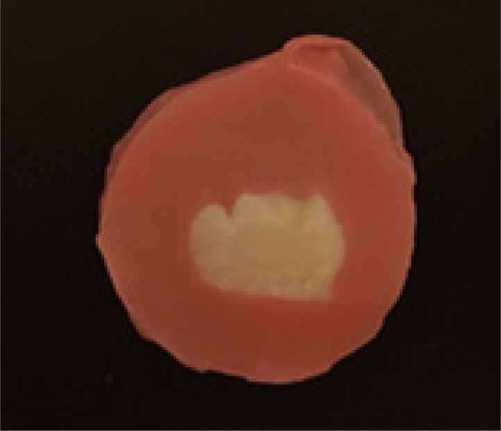
Mesiodistal sectioning of the tooth was done and the tooth was sectioned into two halves using a low-speed diamond cutting blade. One-half of the sectioned teeth were subjected to an immediate subgroup and the other part was used for aging. On the buccal surface of the tooth specimen, a slot was made and these slots were subjected to etching and sealant placement. The sealants were applied according to the manufacturer’s instructions. The first half was tested for immediate microhardness evaluation followed by the other half of the tooth which was tested after aging (Figures 1–3).

Figure 1:
Shows the etched samples on prepared rectangular slots of microtome teeth.

Figure 2:
Shows the 3M™ Clinpro™ Sealant application on the prepared rectangular slots of microtome teeth.

Figure 3:
Shows the Ultra-seal XT Hydro sealant application on the prepared rectangular slots of microtome teeth.
Thermocycling
Aging subgroup samples were subjected to thermocycling, where the samples were placed in a water bath at a temperature of 5 to 55°C for 15 sec at each degree with a dwell duration of 10 sec for 1500 cycles.
Microhardness testing
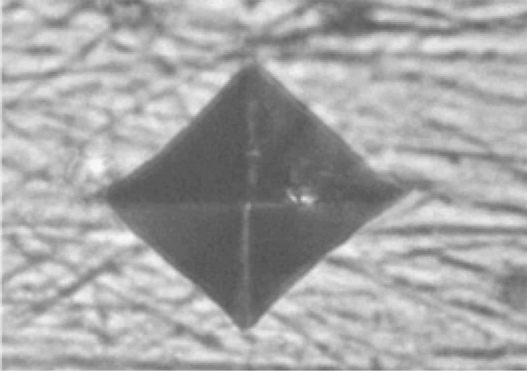
The Microhardness of immediate and aging subgroups for both groups were evaluated using the Vickers hardness instrument. The pyramidal diamond indenter (Figure 4) present in the Vickers hardness tester was used to determine the microhardness of the sealant surface at a 200 g load for 20 sec. All the samples were analyzed and for each point, the Vickers hardness number (VHN) (kg/mm2) was recorded (refer to Figures 5 and 6).

Figure 4:
Shows the Vickers hardness test on the surface of the sample.

Figure 5:
Indenter impression on the tooth surface after treatment with 3M ESPE Clinpro.

Figure 6:
Indenter impression on the tooth surface after treatment with Ultra-seal XT Hydro.
Statistical Analysis
The data was analyzed using SPSS: Statistical Package for Social Sciences(version 23; SPSS Inc., Chicago, Illinois, USA). A p-value of less than 0.05 was considered statistically significant.
Shapiro Wilks test was used to determine the normality of the data, and a non-normal distribution was observed. Hence non-parametric tests were employed. Descriptive statistics were expressed in the form of mean and standard deviation. Mann Whitney U test was used for the comparison of mean microhardness values of Immediate and ageing between Group I and II, and the Wilcoxon signed-rank test was used to compare the mean microhardness values of Immediate and ageing within groups.
RESULTS
Table 1 represents the descriptive data for Immediate and Aging for Groups I and II. The immediate mean microhardness value for Group II (21.06±3.92) was a little higher than Group I (20.08±2.69) and the difference was found to be statistically not significant. A statistically insignificant difference was also observed between the mean microhardness aging values of Group I (28.01±4.02) and Group II (30.04±5.31) using the Man-Whitney U test, but the mean microhardness value for Group II was comparatively higher than the Group I (Table 2). Wilcoxon signed rank test revealed a significant difference within each group for Immediate and Aging time factors (Table 3).
| Outcome | N | Mean | Std. Deviation |
|---|---|---|---|
| Immediate- Group I | 15 | 20.08 | 2.69 |
| Immediate- Group II | 15 | 21.06 | 3.92 |
| Ageing- Group I | 15 | 28.01 | 4.02 |
| Ageing- Group II | 15 | 30.04 | 5.31 |
| Between Groups | Immediate Group I and Group II | Aging Group I and Group II |
|---|---|---|
| Mann whitney U testvalue | 4.50 | 2.81 |
| p value | 0.90 | 0.52 |
| Within Groups | Group I Immediate and Aging | Group II Immediate and Aging |
|---|---|---|
| Wilcoxon test value | 2.08 | 2.02 |
| p value | 0.043* | 0.024* |
DISCUSSION
Two different parameters were used for microhardness evaluation. They are the Knoop hardness number and the Vickers hardness number. Vickers hardness tests were used in numerous cases to determine the material hardness within the microhardness test load range. Vickers hardness test generates a square shape residual indentation which is later measured under a microscope.17
In our study, the microhardness for the sealant Ultra-seal XT Hydro immediately had a mean of 21.06 and after ageing the mean was 30.04 and the difference was statistically significant. Group II ultra seal hydrophilic sealant had a comparatively higher mean aging microhardness value than Group I clinpro conventional sealants. Similar results were observed for hydrophilic sealants in a study done by Sulimany AM et al.,18 where the embrace wet bond group had the highest mean microhardness value compared to other groups.
The indirect measure of micro-hardness will be the Degree of Conversion (DC). The degree of conversion represents the magnitude of a polymer assembled by the monomer conversion after irradiation.19,20 Any low value of degree of conversion in the sealant placed upper and lower layers on occlusal tooth surface may result in poor marginal integrity, which in turn increases dissolution, thereby permitting the cariogenic bacteria proliferation and finally resulting in clinical retention failure of the pit and fissure sealant.21
The present study results were found to be more compared to Mazaheri et al. study results, where they reported a mean micro-hardness of 15.96±4.27 for Fissurit F fluoride-releasing sealant.22 The reason for this lower value compared to the present study results could be attributed to differences in study procedure and assessment tools used for evaluating the outcome parameter.
In a study conducted by Diener et al.,23 Embrace WetBond hydrophilic sealant reported a substantial difference between the means provided by the existing literature and the mean value mentioned in this study, where the result mean was much lower. The difference could be due to the immediate testing of the sample.
In a clinical trial comparing the effectiveness of hydrophilic Embrace WetBond and conventional Helioseal F sealant, two sealants exhibited no significant difference with respect to clinical retention, caries formation and marginal adaptation of sealant.24 The limitations which were observed in the present study were the application of the sealant material on the smooth buccal surface. But in the clinical condition, pit and fissure sealants are placed in various morphological types of pit and fissures with different depths. Another drawback could be in vitro study analysis, which does not provide the same environment as a clinical condition. Hence clinical trials need to be conducted to determine the effectiveness of these sealants.
CONCLUSION
Based on the findings of the present study, Hydrophilic pit and fissure sealants (Group II) had higher micro-hardness mean values compared to conventional sealants (Group I).
Cite this article
Ashwini K, Prabakar J, Ganesh J. Comparative Evaluation of Microhardness of Two Conventional and Hydrophilic Fissure Sealants – An in vitro Study. J Young Pharm. 2023;15(4):725-9.
ABBREVIATIONS
| Std. Deviation | Standard deviation |
|---|
References
- BaniHani A, Deery C, Toumba J, Munyombwe T, Duggal M. The impact of dental caries and its treatment by conventional or biological approaches on the oral health-related quality of life of children and carers. Int J Paediatr Dent. 2018;28(2):266-76. [PubMed] | [CrossRef] | [Google Scholar]
- Souza JGS, Souza SE, Noronha MDS, Ferreira EFE, Martins AMEBL. Impact of untreated dental caries on the daily activities of children. J Public Health Dent. 2018;78(3):197-202. [PubMed] | [CrossRef] | [Google Scholar]
- Jayachandar D, Gurunathan D, Jeevanandan G. Prevalence of early loss of primary molars among children aged 5-10 years in Chennai: A cross-sectional study. J Indian Soc Pedod Prev Dent. 2019;37(2):115-9. [PubMed] | [CrossRef] | [Google Scholar]
- Jayakumar A, Gurunathan D, Mathew MebinG. Correlation of protein level with the severity of early childhood caries – an observational study. J Res Med Dent Sci. 2020;8(7):534-6. [PubMed] | [CrossRef] | [Google Scholar]
- Gandhi JM, Gurunathan D, Doraikannan S, Balasubramaniam A. Oral Health status for primary dentition – A pilot study. J Indian Soc Pedod Prev Dent. 2021;39(4):369-72. [PubMed] | [CrossRef] | [Google Scholar]
- Schwendicke F, Splieth C, Breschi L, Banerjee A, Fontana M, Paris S, et al. When to intervene in the caries process? An expert Delphi consensus statement. Clin Oral Investig. 2019;23(10):3691-703. [PubMed] | [CrossRef] | [Google Scholar]
- Banerjee A, Frencken JE, Schwendicke F, Innes NPT. Contemporary operative caries management: consensus recommendations on minimally invasive caries removal. Br Dent J. 2017;223(3):215-22. [PubMed] | [CrossRef] | [Google Scholar]
- Arora TC, Arora D, Tripathi AM, Yadav G, Saha S, Dhinsa K, et al. An in-vitro evaluation of resin infiltration system and conventional pit and fissure sealant on enamel properties in white spot lesions. J Indian Soc Pedod Prev Dent. 2019;37(2):133-9. [PubMed] | [CrossRef] | [Google Scholar]
- Arslan S, Lipski L, Dubbs K, Elmali F, Ozer F. Effects of different resin sealing therapies on nanoleakage within artificial non-cavitated enamel lesions. Dent Mater J. 2018;37(6):981-7. [PubMed] | [CrossRef] | [Google Scholar]
- Bekes K. Pit and fissure sealants. 2018 [PubMed] | [CrossRef] | [Google Scholar]
- Kühnisch J, Bedir A, Lo YF, Kessler A, Lang T, Mansmann U, et al. Meta-analysis of the longevity of commonly used pit and fissure sealant materials. Dent Mater. 2020;36(5):e158-68. [PubMed] | [CrossRef] | [Google Scholar]
- Prabhakar AR, Murthy SA, Sugandhan S. Comparative evaluation of the length of resin tags, viscosity and microleakage of pit and fissure sealants – an in vitro scanning electron microscope study. Contemp Clin Dent. 2011;2(4):324-30. [PubMed] | [CrossRef] | [Google Scholar]
- Mohapatra S, Prabakar J, Indiran MA, Kumar RP, Sakthi DS. Comparison and Evaluation of the Retention, cariostatic Effect, and Discoloration of Conventional Clinpro 3M ESPE and hydrophilic Ultraseal XT Hydro among 12-15-year-old Schoolchildren for a Period of 6 months: A Single-blind Randomized Clinical Trial. Int J Clin Pediatr Dent. 2020;13(6):688-93. [PubMed] | [CrossRef] | [Google Scholar]
- Prabakar J, Indiran MA, Kumar P, Dooraikannan S, Jeevanandan G. Microleakage assessment of two different pit and fissure sealants: A comparative confocal laser scanning microscopy study. Int J Clin Pediatr Dent. 2020;13(S-1):S29-33. [PubMed] | [CrossRef] | [Google Scholar]
- Güçlü ZA, Dönmez N, Hurt AP, Coleman NJ. Characterisation and microleakage of a new hydrophilic fissure sealant – UltraSeal XT® hydro™. J Appl Oral Sci. 2016;24(4):344-51. [PubMed] | [CrossRef] | [Google Scholar]
- Hahnel S, Henrich A, Bürgers R, Handel G, Rosentritt M. Investigation of mechanical properties of modern dental composites after artificial aging for one year. Oper Dent. 2010;35(4):412-9. [PubMed] | [CrossRef] | [Google Scholar]
- Thakur S, Devadiga D, Hegde NM. Comparative evaluation of the effect of various remineralizing agents on bleached enamel surface: an in vitro study. J Pharm Sci Innov. 2016;4(6):324-8. [CrossRef] | [Google Scholar]
- Sulimany AM, BinSaleh SS, AlYahya E, Bataweel R, Alhussain I, Almahdy A, et al. Effect of aging on the microhardness of different resin-based fluoride-releasing fissure sealants: an in vitro study. J Contemp Dent Pract. 2021;22(10):1144-9. [PubMed] | [CrossRef] | [Google Scholar]
- Borges BC, Bezerra GV, Mesquita A, Pereira MR, Aguiar FH, Santos AJ, et al. Effect of irradiation times on the polymerization depth of contemporary fissure sealants with different opacities. Braz Oral Res. 2011;25(2):135-42. [PubMed] | [CrossRef] | [Google Scholar]
- Silikas N, Eliades G, Watts DC. Light intensity effects on resin-composite degree of conversion and shrinkage strain. Dent Mater. 2000;16(4):292-6. [PubMed] | [CrossRef] | [Google Scholar]
- Kitchens B, Wells M, Tantbirojn D, Versluis A. Depth of cure of sealants polymerized with high-power light emitting diode curing lights. Int J Paediatr Dent. 2015;25(2):79-86. [PubMed] | [CrossRef] | [Google Scholar]
- Mazaheri R, Pishevar L, Keyhanifard N, Ghasemi E. Comparing the effect of topical acidulated phosphate fluoride on micro-hardness of two fissure sealants and one flowable composite. Journal of dental school. Shahid Beheshti University of Medical Sciences. 2019;32(2):103-10. [PubMed] | [CrossRef] | [Google Scholar]
- Diener V, Zinelis S, Eliades T. Water-induced effects on the hardness and modulus of contemporary sealants derived from instrumented indentation testing (IIT). J Contemp Dent Pract. 2019;20(6):653-6. [PubMed] | [CrossRef] | [Google Scholar]
- Askarizadeh N, Heshmat H, Zangeneh N. One-year clinical success of embrace hydrophilic and helioseal-F hydrophobic sealants in permanent first molars: A Clinical Trial. J Dent (Tehran). 2017;14(2):92-9. [PubMed] | [Google Scholar]
