ABSTRACT
ABSTRACT
This case presents a 3-month-old infant with a progressively enlarging Cervical swelling (C2-C3). Magnetic Resonance Imaging revealed a 17×4 mm cystic lesion, consistent with a diagnosis of Cervical Myelomeningocele (cMMC), a rare Neural Tube Defect (NTD) that accounts for only 1-5% of all NTDs. cMMC is characterized by herniation of the spinal cord and meninges through a vertebral defect, often leading to neurological deficits and potential complications such as hydrocephalus and spinal deformities. Genetic predisposition and maternal folic acid deficiency are significant risk factors, with inadequate folic acid intake during early pregnancy being a well-established cause of NTDs. Surgical repair was performed with meticulous dural sac closure and cerebrospinal fluid drainage. The infant demonstrated stable postoperative recovery without immediate complications. This case highlights the importance of maternal folic acid supplementation in preventing NTDs and underscores the need for early diagnosis and surgical intervention to improve outcomes in patients with cMMC.
INTRODUCTION
Myelomeningocele is a severe congenital neural tube defect characterized by the herniation of neural elements and meninges through a vertebral defect, resulting from incomplete closure of the neural tube during embryonic development, typically between 24 and 26 days of gestation (Meyer-Heim et al., 2003). Myelomeningocele most commonly occurs in the lumbosacral or thoracolumbar regions. Cervical myelomeningoceles are rare, comprising only 1-5% of all neural tube defects (Musa and Gots, 2017). The primary risk factors for developing myelomeningocele include genetic predispositions and maternal dietary factors, particularly folic acid deficiency. Although only 5% of cases have a family history, women with an affected family member have a 3-8% increased risk (Ntimbani et al., 2020). Insufficient folic acid during pregnancy is associated with a two to eightfold increase in risk (Trudell and Odibo, 2014). In the U.S., folic acid fortification significantly reduced the incidence of myelomeningocele (Adzick, 2010). Additionally, pre-conception folic acid supplementation has been shown to lower the incidence by up to 70%, especially for high-risk women (Fieggen and Stewart, 2014).
Myelomeningocele is frequently linked to various congenital anomalies, including chromosomal abnormalities such as Trisomy 18 (Edwards syndrome) and Trisomy 13 (Patau syndrome), as well as structural abnormalities like Chiari II malformation, diastematomyelia, syringomyelia, arachnoid cysts, hydrocephalus, and tethering of the spinal cord (Aaronson et al., 2003). Elevated Maternal Alpha-Fetoprotein (MSAFP) is a significant marker in prenatal screening, detected in maternal blood or amniotic fluid, and may suggest the presence of neural tube defects (Jones et al., n.d.) Early diagnosis using prenatal ultrasonography and foetal MRI is critical for identifying the lesion and associated anomalies. Postnatal imaging and surgical intervention are essential for management, especially in cervical cases due to the critical role of the cervical spine in supporting head movement, breathing, and upper limb function (Huang et al., 2010). The management of congenital meningocele and myelomeningocele involves early surgical intervention using standard micro neurosurgical techniques, including the careful removal of the lesion and intradural exploration of the sac to minimize neurological deficits, ensure optimal neural function, and reduce the risk of long-term complications (Hosseini-Siyanaki et al., n.d.) This case underscores the rarity of cervical myelomeningocele, characterized by an open spinal dysraphism at the C2-C3 level with minimal neurological deficits, and highlights the importance of maternal folic acid supplementation in preventing neural tube defects, emphasizing its relevance in pharmacy practice for promoting preconception and prenatal care.
CASE DESCRIPTION
A 3-month-old female, second-born of a consanguineous marriage from a rural area, was delivered via normal vaginal delivery and presented to Dhiraj Hospital with a swelling on the dorsal side of her neck noted soon after birth. The swelling had been progressively increasing in size since birth; it was non-transilluminating, smooth in consistency, and immobile. It is noteworthy that the mother, a homemaker, had not been taking regular folic acid supplements during her pregnancy, a factor associated with neural tube defects, which could have contributed to the patient’s condition.
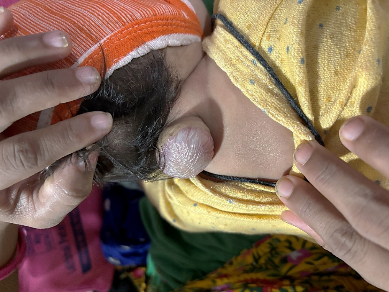
On examination, the patient presented with a swelling over the neck, measuring approximately 4×3 cm with no associated local tenderness, redness, or discharge as shown in Figure 1. However, two days prior to presentation, the swelling became hot to the touch, and the patient developed a mild fever, cough, and itching. In the prenatal period, the pregnancy was booked and routinely followed. After delivery, the patient was admitted to the Neonatal Intensive Care Unit (NICU) for 2-3 days of observation due to neck swelling. Postnatally, the patient had only received the birth doses of immunizations and had been exclusively breastfed.

Figure 1:
Clinical photograph showing a cystic swelling at the nape of the neck, characteristic of cervical myelomeningocele.
The patient’s developmental milestones were largely appropriate for her age. In terms of gross motor skills, she no longer exhibited the Asymmetric Tonic Neck Reflex (ATNR) and showed minimal head lag when pulled to a sitting position. Her head control was improving, though it still occasionally bobbed forward. In ventral suspension, her head was held beyond the plane of her body. Fine motor skills were notable for bringing her hands together at midline during play. Socially, the child recognized her mother and responded to sounds, showing an awareness of her environment.
On physical examination, the patient appeared generally well, without pallor, icterus, cyanosis, clubbing, edema, or lymphadenopathy. The neurological examination was within normal limits. Cranial Nerve (CN) testing showed positive reactions to light (CN II), normal eye movements (CN III, IV, VI), intact corneal and gag reflexes (CN V, IX, X), and normal auditory response (CN VIII). There was no deviation of the mouth or tongue (CN VII, XII). Tone and power in the upper and lower limbs were normal, with muscle power graded 3/5. Examination of the respiratory system revealed clear bilateral air entry, and cardiovascular assessment noted normal heart sounds with no murmurs. No organomegaly was detected on abdominal palpation.
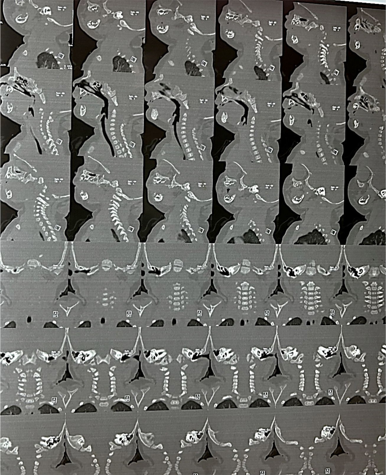
An MRI of the brain was unremarkable, with no evidence of parenchymal lesions, hydrocephalus, or acute infarction. However, MRI of the cervical spine revealed a 17×4 mm cystic lesion at the C2-C3 level, consistent with an open spinal dysraphism. The lesion, characterized by septae-like structures suggesting the presence of neuronal components, caused focal posterior tenting of the cervical spinal cord. These findings were diagnostic of a cervical myelomeningocele, a type of neural tube defect where the spinal cord and meninges protrude through a defect in the vertebral column. Sagittal and Coronal Computed Tomography images of the cervical spine show a bony defect at C2-C3 with posterior herniation of neural structures, while ruling out spondylolysis, spondylolisthesis, and soft tissue abnormalities; atlanto-axial and atlanto-occipital joints appear unremarkable (as shown in Figure 2). Blood tests showed prolonged prothrombin time (19 sec), elevated activated partial thromboplastin time (37.2 sec), low haemoglobin, and elevated neutrophils, lymphocytes, eosinophils, and platelets, requiring close monitoring of coagulation status for surgery.

Figure 2:
Sagittal and coronal CT images of the cervical spine showing a bony defect at C2-C3 with posterior herniation of neural structures.
The patient was initiated on supportive medical management with Vitanova Drops (0.5 cc Once Daily) and Rxplus Oral Drops (0.5 cc OD) to address potential nutritional deficiencies commonly seen in neonates with neural tube defects, along with an Intramuscular (IM) injection of Vitamin K (1 mg) to prevent haemorrhagic disease of the newborn, as such patients are at increased risk of coagulopathy. Given the diagnosis of cervical myelomeningocele-a serious congenital anomaly associated with risks of infection, neurological deterioration, and cerebrospinal fluid leakage-surgical intervention was warranted. On the fourth day of admission, the patient underwent myelomeningocele repair. Preoperative medications included Intravenous (IV) ondansetron (0.5 mg) to prevent postoperative nausea and vomiting, midazolam (0.1 mg IV) for sedation and anxiolysis, paracetamol (50 mg IV) for analgesia and antipyresis, glycopyrrolate (0.02 mg IV) to reduce secretions and prevent bradycardia, ceftriaxone (200 mg IV) for prophylactic antibiotic coverage, and fentanyl for intraoperative pain management. General anaesthesia was induced using propofol, a rapid-onset hypnotic agent, and succinylcholine, a depolarizing neuromuscular blocker, to facilitate endotracheal intubation and ensure adequate muscle relaxation during surgery.
During the procedure, a skin incision was made around the meningocele sac, which was deepened to expose the dural sac. The meningocele sac was incised, and Cerebrospinal Fluid (CSF) was drained. The dural sac was then meticulously closed in layers, with irrigation to ensure a clean operative field. The procedure was completed successfully, and the patient was given a reversal of anaesthesia with neostigmine and glycopyrrolate.
Post-operatively, the patient was closely monitored, and aminoglycosides were withheld for 24 hours to reduce the risk of nephrotoxicity. Zostum IV (Piperacillin+Tazobactam) and Amikacin IV were administered for broad-spectrum antibiotic coverage. Pantoprazole IV was given to prevent stress ulcers, Emset IV (Ondansetron) for nausea control, and Febrinyl IV (Paracetamol) for pain and fever management. Combimist nebulization (Ipratropium+Salbutamol) was provided as per paediatric dosing to support airway function and ease breathing.
This case underscores the significance of early detection and timely surgical management in congenital spinal anomalies such as cervical myelomeningocele. The mother’s irregular intake of folic acid during pregnancy may have contributed to the development of this neural tube defect. The successful surgical repair of the myelomeningocele highlights the importance of a multidisciplinary approach, particularly in rural settings where access to specialized care may be limited. The patient’s prognosis following surgery was positive, and she continues to be monitored for further developmental milestones and any potential complications.
DISCUSSION
Cervical myelomeningocele is a rare neural tube defect causing spinal cord and meningeal herniation, usually in the lumbar or sacral regions, with cervical cases being exceptionally rare. A critical factor in the prevention of NTDs is the adequate intake of folic acid during the preconception and early pregnancy period. The mother in this case, however, was not taking regular folic acid supplements during pregnancy, which could have contributed to the occurrence of the anomaly. This emphasizes the need for public health interventions in rural areas to ensure that women of childbearing age receive proper folic acid supplementation and prenatal care.
In this case, the swelling on the dorsal side of the neck, present since birth and progressively increasing in size, was the initial sign that led to the discovery of the cervical myelomeningocele. While the patient presented with no neurological deficits, this condition can potentially lead to complications such as hydrocephalus, motor deficits, and respiratory issues due to the involvement of the upper cervical spine. Fortunately, the MRI findings confirmed that there was no hydrocephalus, and the child’s brain development appeared normal. The presence of a septate cyst and the posterior tenting of the cervical spinal cord were crucial diagnostic indicators for the myelomeningocele. Early surgical intervention was essential to prevent further complications, and the patient’s surgery was performed successfully without post-operative complications.
The management of cervical myelomeningocele involves prompt surgical repair, which reduces the risk of infection, particularly meningitis, and prevents further neurological damage (Sydney Children’s Hospitals Network, 2011). In this case, the surgical approach involved a meticulous incision around the meningocele sac, drainage of Cerebrospinal Fluid (CSF), and a layered closure of the dural sac. This method is standard in cases of myelomeningocele repair and aims to protect the exposed neural tissue while preventing leakage of CSF, which could lead to complications such as CSF fistula or meningitis.
When comparing this case to others in the literature, cervical myelomeningocele remains an exceedingly rare diagnosis. In contrast to the case reported by Armas-Melián et al., (2020), where the newborn with cervical myelomeningocele at the C1-C3 level presented with severe complications such as hydrocephalus and significant neurological deficits, including quadriparesis, our patient exhibited no major neurological deficits apart from mildly reduced muscle power (3/5) in the limbs, with no other complications. Other cases include an 11-month-old who had postoperative cardiorespiratory arrest but was resuscitated (Sriharsha et al., 2020), and an 18-year-old with untreated cMMC developing leiomyosarcoma (Mohindra et al., 2021). This suggests that patients with cervical myelomeningocele may be at risk for spinal deformities and will need ongoing follow-up to assess for late complications.
Studies suggest that early surgical intervention for myelomeningocele can lead to favorable outcomes. For instance, a study reported that early and meticulous surgical repair resulted in low complication rates, with neurological deficits observed in only 2.5% of cases (Shah and Shroff, 2019). Additionally, research indicates that prenatal surgery for myelomeningocele may not fully prevent long-term motor deficits, highlighting the complexity of treatment timing (Ben Miled et al., 2020). Therefore, while early surgical intervention is generally associated with improved motor outcomes, the optimal timing and approach may vary based on individual circumstances. The absence of hydrocephalus in cervical cases, which was also noted in our patient, further contributed to better prognoses.
Insufficient folic acid intake during pregnancy is a modifiable risk factor for Neural Tube Defects (NTDs). In India, studies have estimated the pooled birth prevalence of NTDs to be between 4.1 and 4.5 per 1,000 births (Bhide, 2021). A 2017 survey in Haryana revealed that a significant proportion of women of reproductive age had low folate and vitamin B12 levels, highlighting the need for improved prenatal care (Das et al., 2021). This underscores the importance of enhancing public health initiatives in rural areas to educate women about the benefits of periconceptional folic acid supplementation in preventing NTDs.
CONCLUSION
In conclusion, this case of cervical myelomeningocele underscores the importance of early detection, timely surgical intervention, and ongoing post-operative care. Comparing this case to similar cases in the literature reveals that, despite the rarity of cervical myelomeningocele, outcomes are generally favourable when treated early. However, ongoing monitoring is essential to address any potential late complications, such as spinal deformities or motor deficits. This case also highlights the critical role of folic acid supplementation in preventing NTDs and calls for enhanced efforts in public health education and prenatal care, especially in rural and consanguineous populations.
Cite this article:
Patel M, Gohil D. Cervical Myelomeningocele: A Rare Neural Tube Defect: A Case Report. J Young Pharm. 2025;17(3):747-51.
ACKNOWLEDGEMENT
The authors thank Dr. Vikrant Keshari, Neurosurgeon at Dhiraj Hospital, along with the hospital staff and the Department of Pharmacy, Sumandeep Vidyapeeth (Deemed to be University), for their support and expert guidance.
ABBREVIATIONS
| CMMC | Cervical Myelomeningocele |
|---|---|
| NTD | Neural Tube Defect |
| CNS | Central Nervous System |
| MRI | Magnetic Resonance Imaging |
| CSF | Cerebrospinal Fluid |
| IV | Intravenous |
| CN | Cranial Nerve |
| OD | Once Daily. |
References
- Aaronson O. S., Hernanz-Schulman M., Bruner J. P., Reed G. W., Tulipan N. B.. (2003) Myelomeningocele: Prenatal evaluation-Comparison between transabdominal US and MR imaging. Radiology 227: 839-843 https://doi.org/10.1148/radiol.2273020535 | Google Scholar
- Adzick N. S.. (2010) Fetal myelomeningocele: Natural history, pathophysiology, and in-utero intervention. Seminars in Fetal and Neonatal Medicine 15: 9-14 https://doi.org/10.1016/j.siny.2009.05.002 | Google Scholar
- Armas-Melián K., Iglesias S., Ros B., Martínez-León M. I., Arráez M. Á.. (2020) Cervical myelomeningocele with CSF leakage: A case-based review. Child’s Nervous System 36: 2615-2620 https://doi.org/10.1007/s00381-020-04743-y | Google Scholar
- Ben Miled S., Loeuillet L., Duong Van Huyen J.-P., Bessières B., Sekour A., Leroy B., Tantau J., Adle-Biassette H., Salhi H., Bonnière-Darcy M., Tessier A., Martinovic J., Causeret F., Bruneau J., Saillour Y., James S., Ville Y., Attie-Bitach T., Encha-Razavi F., Stirnemann J., et al. (2020) Severe and progressive neuronal loss in myelomeningocele begins before 16 weeks of pregnancy. American Journal of Obstetrics and Gynecology 223: 256.e1–256.e9 https://doi.org/10.1016/j.ajog.2020.02.052 | Google Scholar
- Bhide P., Kar A.. (2021) Birth defects in India : 235-249 https://doi.org/10.1007/978-981-16-1554-2_10 | Google Scholar
- Das R., Duggal M., Rosenthal J. (2021) Folate and vitamin B12 status in women of reproductive age in rural Haryana, India: Estimating population-based prevalence for neural tube defects. Public Health Nutrition 24: 450-457 https://doi.org/10.1007/978-981-16-1554-2_10 | Google Scholar
- Fieggen K., Stewart C.. (2014) Aetiology and antenatal diagnosis of spina bifida. South African Medical Journal 104: 218-221 https://doi.org/10.7196/SAMJ.8039 | Google Scholar
- Hosseini-Siyanaki M.-R., Liu S., Dagra A., Reddy R., Reddy A., Carpenter S. L., Khan M., Lucke-Wold B., et al. (2024) Surgical management of myelomeningocele. Neonatal 4 https://doi.org/10.35702/neo.10008 | Google Scholar
- Huang S.-L., Shi W., Zhang L.-G.. (2010) Characteristics and surgery of cervical myelomeningocele. Child’s Nervous System 26: 87-91 https://doi.org/10.1007/s00381-009-0975-7 | Google Scholar
- Jones J., Campos A., Elfeky M. (n.d.) Myelomeningocele. Radiopaedia.org. https://doi.org/10.53347/rID-5791 | Google Scholar
- Meyer-Heim A. D., Klein A., Boltshauser E.. (2003) Cervical myelomeningocele: Follow-up of five patients. European Journal of Paediatric Neurology 7: 407-412 https://doi.org/10.1016/s1090-3798(03)00108-9 | Google Scholar
- Mohindra S., Batish A., Tripathi M., Nambiyar K., Gupta K.. (2021) Leiomyosarcoma in a cervical myelomeningocele: A rare complication in a neglected case. Child’s Nervous System 37: 2097-2103 https://doi.org/10.1007/s00381-020-04928-5 | Google Scholar
- Musa G., Gots A.. (2017) Cervical meningomyelocele: A case report and review of the literature. Medical Journal of Zambia 44: 203-206 https://doi.org/10.1007/s00381-020-04928-5 | Google Scholar
- Ntimbani J., Kelly A., Lekgwara P.. (2020) Myelomeningocele: A literature review. Interdisciplinary Neurosurgery 19: Article 100502 https://doi.org/10.1016/j.inat.2019.100502 | Google Scholar
- Shah A. S., Shroff M.. (2019) Analysis of the short-term outcomes of surgical repair of myelomeningocele. Journal of Neurosurgical Sciences 63: 1-8 https://doi.org/10.1016/j.inat.2019.100502 | Google Scholar
- Sriharsha R., Kataria K. K., Meena S., Jangra K., Bloria S.. (2020) Postoperative cardiorespiratory arrest in a case of cervical meningocele. Surgical Neurology International 11: 45 https://doi.org/10.25259/SNI_461_2019 | Google Scholar
- Sydney Children’s Hospitals Network (SCHN). (2011) Myelomeningocele in neonates: Pre and post-operative – GCNC CHW. https://doi.org/10.25259/SNI_461_2019 | Google Scholar
- Trudell A. S., Odibo A. O.. (2014) Diagnosis of spina bifida on ultrasound: Always termination? Best Practice and Research. Clinical Obstetrics and Gynaecology 28: 367-377 https://doi.org/10.1016/j.bpobgyn.2013.10.006 | Google Scholar
