ABSTRACT
Presenting case report of necrotizing fasciitis, a rare bacterial infection, bacteria grow in the superficial layer of the fascia and release toxins, causing the infection to spread through the fascia. This is a serious disease that spreads quickly and requires immediate treatment. Compartment syndrome is one of the complications of necrotizing fasciitis. The prognosis is reliable based on clinical examination and assessment of intracompartmental pressure. Severe pain is important sign of this. Either the relatively rigid and closed muscle space, which is enclosed by fascia and bone, experiences bleeding and oedema. The case is unique in terms of the misdiagnosis and complication of the disease. Patient ageing forty-four years came to an emergency room with the complaint of pain, redness and severe swelling in right leg in the last 7 days. He went to a local hospital a week ago where cellulitis was diagnosed but before 2 days the swelling and pain got severe and so he came in as an emergency. It was noted here that he was misdiagnosed by cellulitis and so Laboratory Risk Indicator for Necrotizing Fasciitis (LRINEC) score was performed which revealed the intermediate risk of necrotizing Fasciitis. He was immediately admitted to operation room for surgical procedure called debridement with fasciotomy, after the surgery antibiotic treatment started and was continued for a week. Along with this, dressing of the surgical site was performed. Plastic surgery was done at surgical site after 15 days. With this treatment the recovery was good and the therapeutic goals were all achieved. So, it is better that this type of diagnosis should be identify at stage-1 to decrease the mortality rate.
INTRODUCTION
Tissues dying is referred to as necrotizing. Inflammation of the fascia is referred to as fasciitis. Life-threatening illness known as Necrotizing fasciitis was initially identified by joseph jones during the American war, who stated that the “affected part of skin melts away”.1 A flesh-eating bacterial condition called necrotizing fasciitis. Necrotizing fasciitis is most usually brought on by Group A Streptococcus (GAS) bacteria. But several other types of bacteria also cause this disease. A layer of connecting tissue below the skin called fascia usually gets infected by these bacteria leading to this disease. Typically, cuts, burns, surgical wounds, puncture wounds, and insect bites allow bacteria to enter the body.2 A combination of enzymes, endotoxins, and exotoxins are released by bacterial development within the superficial fascia, causing infection to propagate across this fascia. Poor microcirculation, ischemia, and ultimately cell death and necrosis result from this process in the affected tissues.3 Patient is at high risk of necrotizing fasciitis if he has one of the following disease conditions: diabetes, cancer, heart disease, lung disease, kidney disease, steroid use etc. Most common site of infection is the upper limb and lower limb extremities, trunk, head and neck.2 Painful disease known as compartment syndrome is caused by ICP (Increased Compartment Pressure) within a closed osteofascial compartment. It is both short-term and long-term. A surgical emergency, Acute Compartment Syndrome (ACS) can lead to soft tissue necrosis and irreversible impairment if the pressure is not promptly removed.4
CASE PRESENTATION
A forty four-year-old man of Dravidian race who had been complaining about blisters, redness and severe swelling in his right leg (Figure 1) for the last 5 days appeared in the emergency room. The patient recalled that 7 days ago he had a minor accident and fell from his bike (Figure 2). Due to that accident, he got a minor injury on his right leg, after 2 days he observed redness, pain, and swelling with a fever. He went to a local hospital. A primary diagnosis of cellulitis was made and he was started on a high dose of antibiotic (zostum). After 2 days situation get worse with progressive severe swelling, and he came to tertiary care for further evaluation and management. Physical examination revealed multiple superficial wounds. He appeared ill. The whole leg was swollen and it was warm and tender to the touch. Their blood pressure was normal. Doctor decided to perform LRINEC (Laboratory Risk Indicator for Necrotizing Fasciitis) score. In this Hematological examination revealed a white blood count of 23000 (normal range 4000-11000/cm), hemoglobin 8 g/dl (normal value 14.5-18 g/dl), and CRP 161 mg/L (normal value less than 6 mg/L).2 The other two parameter, glucose and sodium levels were normal. From the examination diagnosis of necrotizing fasciitis with compartment syndrome was made. Surgical debridement with fasciotomy (Figure 3) was carried out urgently and Inj. zostum 3 gm, I.V once a day was continued with Inj. Amikacin (250mg/mL Twice daily), Inj. Pantoprazole (40 mg Twice daily), and Inj. Periset (2mg/mL Twice daily). During debridement, swab culture has taken. Examination revealed hemoglobin 8 gm, red blood cell 4.23 mL/cm, white blood count 19300/cm, platelet count 192000/cm, neutrophils 80%, lymphocyte 10%, CRP 136 mg/L. The swab culture showed no pathogenic organisms. The pain, swelling and intercompartmental pressure was reduced and there were no fresh complaints and the patient was vitally stable. Later the patient was transferred to the hospital for skin grafting surgery (Figure 4) for the management of the fasciotomy wound. By following this treatment, the recovery was good and there was no disease progression after surgery. The patient almost recovered in three months (Figure 5).
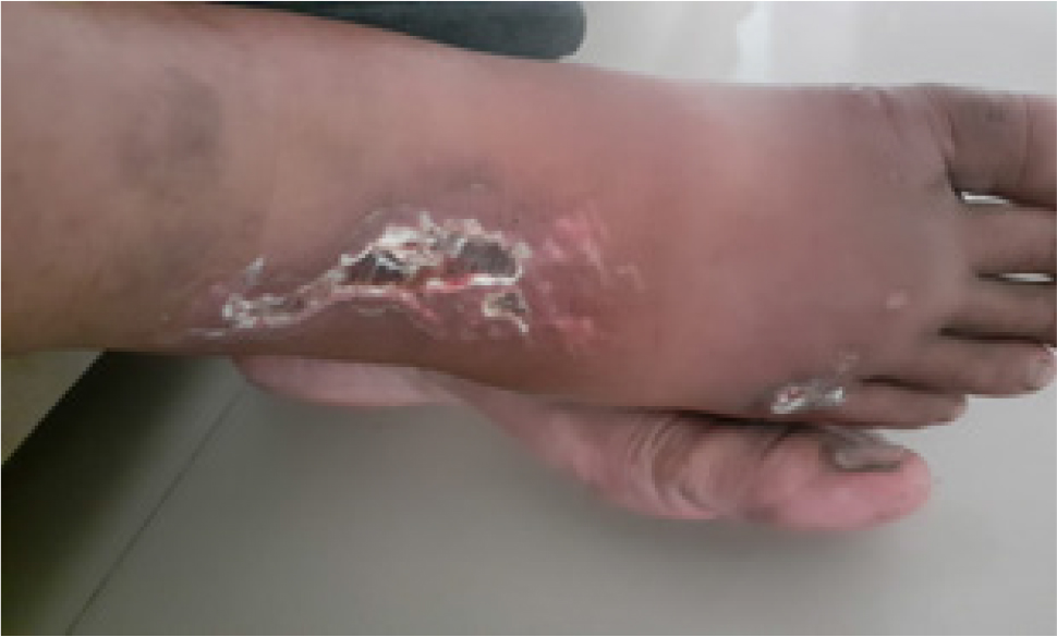
Figure 1:
Severe swelling in right leg.
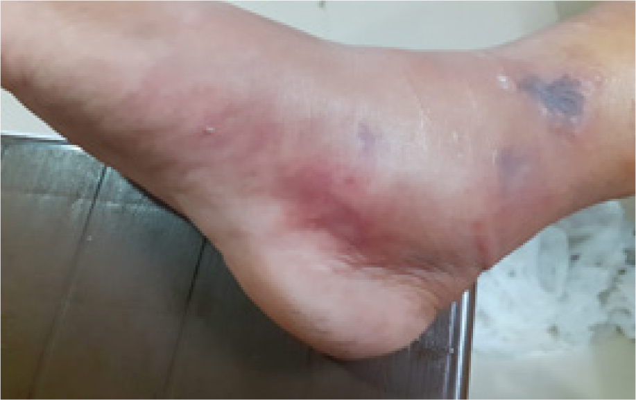
Figure 2:
Photo of patient’s right leg after he fell.
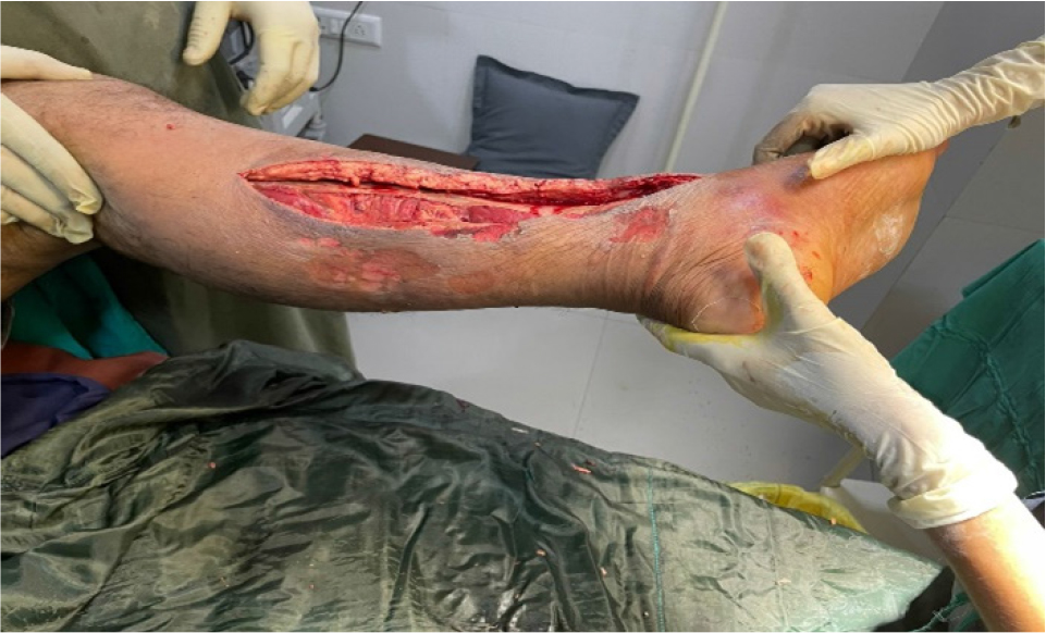
Figure 3:
After debridement with fasciotomy.
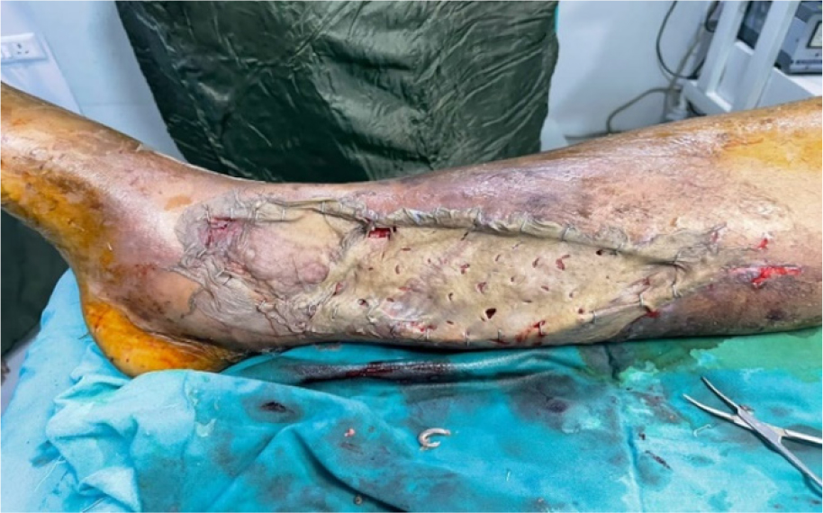
Figure 4:
After skin grafting surgery.
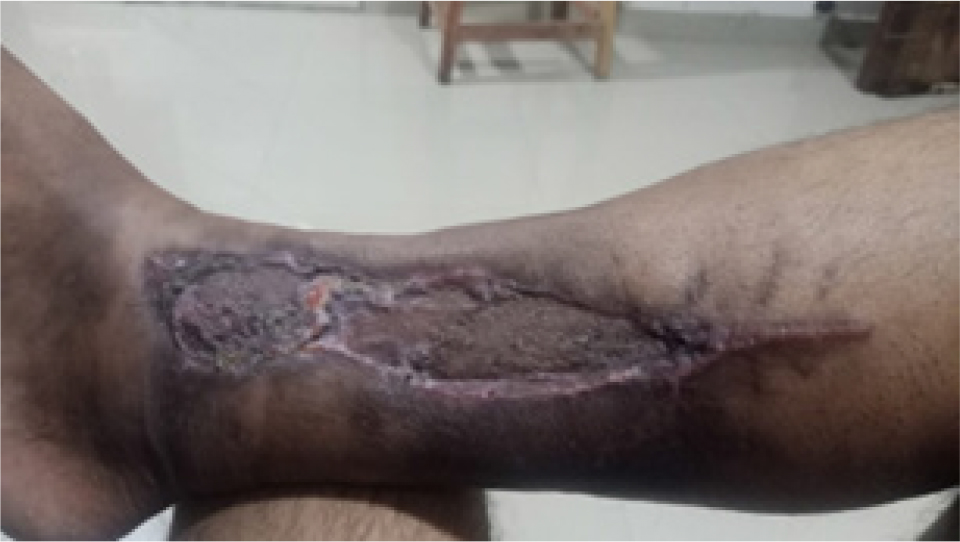
Figure 5:
Recent photo of patient’s right leg.
DISCUSSION
A contagious illness, NF is characterized by fast bacterial spread along the fascial planes. It as an uncommon disease, with an incidence of 1.5 cases per 100,000 people annually, although the mortality rate is relatively significant, at 20-30%. NF typically manifest in the upper or lower extremities of patients (approximately 57.8%), however occurrences of cervical or abdominal NF have also been reported.5 most commonly necrotizing fasciitis caused by group of streptococcus bacteria. In this case patient had received high dose of antibiotic zostum before swab culture so no pathogenic organism found in it and one might speculate that this organism was inhibited by the antibiotic agent. In order to distinguish necrotizing fasciitis from severe cellulitis or abscess, the LRINEC was established. The phenomenon was first described by Wong C. et al. This is based on six typical serum measures, including creatinine, CRP, Hb, WBC, serum sodium and glucose. Necrotizing fasciitis is more likely if the LRINEC is less than six.6 In our patient the score is 7 that indicate that patient is at intermediate risk of having NF. The main course of treatment for NF is early, vigorous surgical exploration and debridement of necrotic tissue. An effective broad-spectrum parenteral antibiotic regimen is administered alongside the surgery. The mainstay of NF treatment is surgical debridement, which has a significantly lower mortality rate than waiting even a few hours for surgery.7,8
CONCLUSION
In this case report we accentuate on a 44-year-old male patient who has necrotizing fasciitis, a rare condition with a poor prognosis. It is typically misdiagnosed with other soft tissue infections. Just like in this case it was misdiagnosed with cellulitis, so physical examination should be performed accurately. Best way to distinguish necrotizing fasciitis from cellulitis and for accurate diagnosis, the LRINEC score is used. Physicians must recognize necrotizing fasciitis as soon as possible, since early diagnosis of infection has great impact on mortality rate and improve the quality of life of the patient.
References
- . Committee on Infectious Diseases. Severe invasive group A streptococcal infections: a subject review. Pediatrics. 1998;101(1):136-40. [Google Scholar]
- Sarani B, Strong M, Pascual J, Schwab CW. Necrotising fasciitis: current concepts and review of the literature. J Am Coll Surg. 2009;208(2):279-88. [Google Scholar]
- Schwartz JT, Brumback RJ, Lakatos R, Poka A, Bathon GH, Burgess AR, et al. Acute compartment syndrome of the thigh-a spectrum of Inj.ury. J Bone Joint Surg Am. 1989;71(3):392-400. [Google Scholar]
- Davies HD, mcgeer A, Schwartz B, Green K, Cann D, Simor AE, et al. Invasive group A streptococcal infections in Ontario, Canada. Ontario Group A streptococcal study group. N Engl J Med. 1996;335(8):547-54. [PubMed] | [CrossRef] | [Google Scholar]
- Wong CH, Khin LW, Heng KS. The LRINEC (Laboratory Risk Indicator for Necrotizing Fasciitis) score: a tool for distinguishing necrotizing fasciitis from other soft tissue infections. Crit Care Med. 2004;32(7):1535-41. [PubMed] | [CrossRef] | [Google Scholar]
- [PubMed] | [CrossRef] | [Google Scholar]
- Mchenry CR, Piotrowski JJ, Petrinic D, Malangoni MA. Determinants of mortality for necrotizing soft-tissue infections. Ann Surg.. 1995;221(5):558-63. [PubMed] | [CrossRef] | [Google Scholar]
