ABSTRACT
Background
The efficacy of the anticancer agent palbociclib can be limited by its conventional release profile. Developing nano-formulations presents a viable approach to modulate its delivery for improved therapeutic outcomes. This work aimed to design, optimize, and characterize chitosan-based Polymeric Nanoparticles (PNs) for the oral delivery of palbociclib, with a specific focus on controlling particle size and surface charge.
Materials and Methods
Palbociclib-loaded PNs were fabricated using a solvent evaporation technique. A Central Composite Design was employed to optimize the concentrations of chitosan and Polyvinyl Alcohol (PVA). The lead formulation was thoroughly characterized using FT-IR, SEM, and in vitro release studies, with its release kinetics modelled.
Results
An optimized formulation was identified with a particle size of 337.8 nm and a zeta potential of 32.4 mV, which aligned closely with statistical predictions. The system exhibited high drug entrapment (81.28%) and provided a sustained release profile, with 90% of the drug released over 26.1 hr following a diffusion-erosion mechanism. This was a substantial extension over the rapid release of the pure drug.
Conclusion
The findings confirm the successful development of a chitosan-based nanoparticle system capable of offering sustained release of palbociclib, highlighting its significant potential for enhancing oral drug delivery in cancer treatment.
INTRODUCTION
Nanotechnology remains an enigmatic concept for many individuals, yet it is already shaping our future and promises to have a profound impact when combined with biotechnology and information technology (Chakrabortyet al., 2025; Martin, 2000). Nanocapsules and nanospheres are included in the term nanoparticle and these two are identical with the exception in their morphology (Schaffazicket al., 2003). Polymeric Nanoparticles (PNPs) have the ability of encapsulating various therapeutic agents, increase bioavailability, and produce controlled release which in turn, increase therapeutic efficacy and decrease the side effects (Aldaais, 2018, Amanzioet al., 2020). Polymer characterization used in the production of PNPs determines directly the physicochemical nature of PNPs including their size, shape, surface charge and drug-loading capacity (Luet al., 2021). One factor that contributes significantly to the effectiveness of PNPs in targeted drug delivery is the capability of crossing biological barriers and the capacity to release the cargo at a specific location where it is supposed to act. This requires an in-depth understanding of the physical characteristics of the polymer, its molecular weight, hydrophilicity/hydrophilicity, and its fictionalization, which all influence considerably the interaction of PNPs with a biological environment (Fanget al., 2011). The technique of formulation and characterization is imperative to the performance of nanoparticle-based drug delivery systems owing to the fact that it does influence physicochemical properties of the drug delivery system that indirectly do affect therapy outcomes, targeting capabilities, and drug delivery effectiveness. Assessing the size, the shape, the surface charge and related particularities of the nanoparticles is critical in achieving maximum effectiveness of nanoparticles that is exercised in the concept of drugs delivery. The process ensures that the nanoparticles are tailored to meet specific therapeutic needs which in turn make them more efficient in delivering medicines where they are needed (Kankalaet al., 2021).
Pfizer came up with the drug palbociclib that will be used to treat HER2-negative and HR-positive breast cancer. It specifically suppresses two cyclin-dependent kinases, i.e., CDK4 and CDK6. Palbociclib was the first CDK4/6 to be approved as cancer treatment. Due to low water solubility and because of first-pass metabolism it also does not have a good oral bioavailability at 46%. Polymaer based nanoparticle can thus be regarded as an acceptable drug delivery system by enhancing poor oral bioavailability of palbociclib (Roccaet al., 2017, Patra, 2018). Hence, the present study is planned to design, optimize, and characterize chitosan-based polymeric Nanoparticles (PNs) for the oral delivery of palbociclib, with an emphasis on achieving controlled particle size and surface charge.
MATERIALS AND METHODS
Materials
Palbociclib was generously provided by Natco Pharma, Hyderabad. Chitosan, Acetone, Ethanol and Polyvinyl Alcohol were perches from Loba Chemie Pvt. Ltd., Mumbai, Maharashtra, India. All Chemicals and API were passed for analytical grade.
Pre-formulation study
Preformulation studies analyze the drug’s molecular and physical properties, both alone and with excipients, to guide effective and scalable formulation design (Krishnakumaret al., 2011).
Drug-Excipients Interactions by Fourier Transform Infra-Red (FTIR) spectroscopy
FT-IR analysis (4000-400 cm⁻¹) was performed on palbociclib, chitosan, PVA, their physical mixtures, and lyophilized formulations (with/without drug) using a Bruker Alpha II spectrometer (1:1 sample ratio) (Krishnakumaret al., 2011).
Formulation of Palbociclib polymers-based nanoparticles
To prepare PNPs, the solvent evaporation technique was used. Chitosan was first dissolved in a solution of 1% acetic acid, and folic acid was then added and stirred for 2 hr. Palbociclib was dissolved in acetone, and a 2% PVA aqueous solution was made by heating water. This organic phase was sonicated into the PVA solution to create an emulsion, while folic acid and 0.3% sodium Tripolyphosphate (TDD) were added as a cross-linking agent. The solvent was evaporated with continuous stirring, and the resulting mixture was centrifuged at 15,000 rpm for 30 min. The final pellet was washed with distilled water to complete the process (Sartajet al., 2021).
Evaluation of Palbociclib Polymeric Nanoparticles
The particle size and zeta potential of palbociclib-loaded nanoparticles were measured using a Zetasizer Nano-ZS (Malvern, UK) with a 633 nm, Data analysis was performed using automated software, based on electrophoretic mobility and the Smoluchowski model (Yanget al., 2010).
PNPs were centrifuged, and the supernatant was analyzed at 261 nm. The precipitate was filtered, washed, weighed, and drug loading was calculated (Liuet al., 2010).
PNPs were centrifuged, and the supernatant was analyzed at 261 nm. Entrapment efficiency was calculated from the free drug content (Mandalet al., 2013).
In vitro Drug Release Studies
The drug release of Palbociclib-loaded nanoparticles and free palbociclib was studied using the dialysis bag method. A 20 mL sample was placed in dialysis bags, immersed in 150 mL of 7.4 pH phosphate buffer at room temperature with constant stirring. At set intervals, 2 mL was withdrawn and replaced with fresh buffer, then analyzed at 267 nm using a UV spectrophotometer (Yuet al., 2016, Xieet al., 2008).
In vitro Drug Release Kinetics
Scanning Electron Microscope (SEM)
SEM (ESEM EDAX XL-30) was used to characterize polymeric nanoparticles. Freeze-dried samples were stored in a gold-coated graphite sample container. The chamber was maintained at 0.6 mm Hg pressure and 20 KV voltages. Scanning electron photomicrographs were taken at various magnifications (Yuanet al., 2012).
RESULTS
Physiochemical properties of palbociclib
Palbociclib is a yellow, amorphous, and odorless powder. It is soluble in ethanol but practically insoluble in water, and exhibits limited solubility in dichloromethane and chloroform.
Preparation of the Standard curve of Palbociclib
A standard calibration curve of Palbociclib was constructed by plotting absorbance against concentration in the range of 5-40 µg/mL. A linear relationship was obtained with a regression coefficient (R²=0.997), confirming adherence to Beer-Lambert’s law (Figure 1).
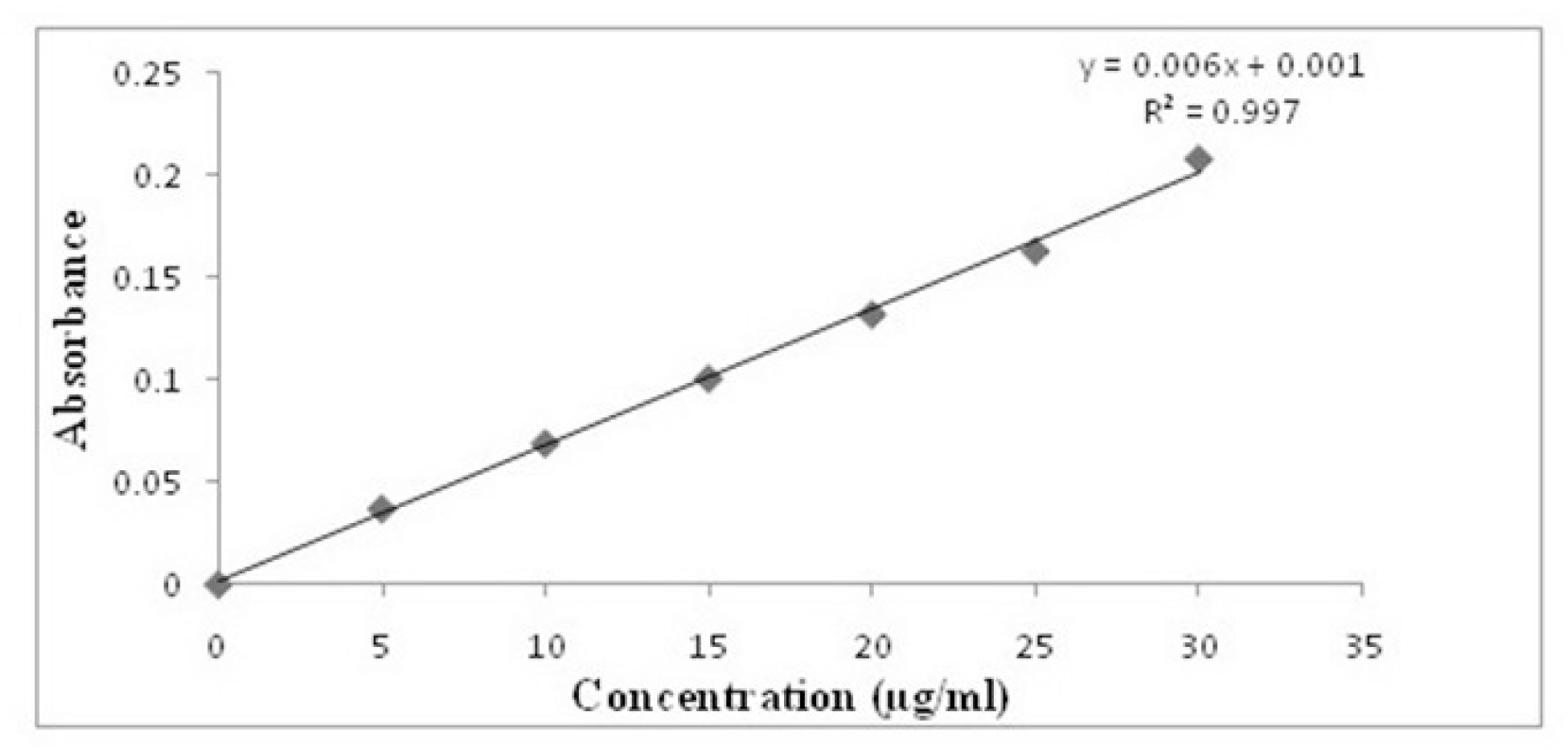
Figure 1:
Graph Plot of Standard Curve of Palbociclib.
Drug Excipients Interaction
Fourier Transform Infrared (FT-IR) spectroscopy was performed to assess potential drug-excipient interactions. Comparative spectra of Palbociclib and its physical mixture with excipients (Figures 2 and 3; Table 1) showed no significant shifts or disappearance of characteristic peaks, indicating the absence of major chemical interactions and confirming the compatibility of Palbociclib with the selected excipients.

Figure 2:
FT-IR spectrums of palbociclib.

Figure 3:
FT-IR spectrums of Palbociclib with the excipients.
| Sl. No. | Wavelength Range | Concentration (µg/mL) | I | II | III | Absorption(nm) (Mean) |
|---|---|---|---|---|---|---|
| 0 | 261 nm | 0 | 0 | 0 | 0 | 0 |
| 1 | 5 | 0.037 | 0.036 | 0.038 | 0.037 | |
| 2 | 10 | 0.069 | 0.07 | 0.068 | 0.069 | |
| 3 | 15 | 0.099 | 0.101 | 0.102 | 0.100 | |
| 4 | 20 | 0.132 | 0.131 | 0.133 | 0.132 | |
| 5 | 25 | 0.163 | 0.162 | 0.164 | 0.163 | |
| 6 | 30 | 0.208 | 0.209 | 0.207 | 0.208 |
Design of Experiments
A statistical design of experiments approach was applied to evaluate the influence of formulation variables on particle size and zeta potential of the prepared chitosan-based Polymeric Nanoparticles (PNs). The evaluated formulations were subjected to particle size and surface charge analysis (Table 2).
| Run | Factor A: Chitosan (mg) | Factor C: PVA (mg) | Folic Acid (mg) | Acetone 80 (mL) | Acetic Acid (mL) | Dist. H2O (mL) | Response 1: MPS (nm) | Response 2: ZP (mV) | ||
|---|---|---|---|---|---|---|---|---|---|---|
| F1 | 100 | 15 | 10 | 5 | 10 | Qs | 402 | 34.5 | ||
| F2 | 60 | 15 | 10 | 5 | 10 | Qs | 376 | 33.47 | ||
| F3 | 20 | 10 | 10 | 5 | 10 | Qs | 412 | 36.76 | ||
| F4 | 20 | 15 | 10 | 5 | 10 | Qs | 398 | 35.65 | ||
| F5 | 60 | 15 | 10 | 5 | 10 | Qs | 389 | 34.67 | ||
| F6 | 60 | 15 | 10 | 5 | 10 | Qs | 382 | 36.65 | ||
| F7 | 20 | 20 | 10 | 5 | 10 | Qs | 411 | 32.41 | ||
| F8 | 60 | 10 | 10 | 5 | 10 | Qs | 401 | 31.2 | ||
| F9 | 100 | 10 | 10 | 5 | 10 | Qs | 412 | 29.87 | ||
| F10 | 60 | 20 | 10 | 5 | 10 | Qs | 389 | 30.94 | ||
| F11 | 100 | 15 | 10 | 5 | 10 | Qs | 399 | 32.12 | ||
| Independent and Dependant variables in CCD | ||||||||||
| Independent Variables | Levels | |||||||||
| Variable | Name | Unit | Low (-1) | High (+1) | ||||||
| A | Chitosan | mg | 100 | 200 | ||||||
| B | Polyvinyl Alcohol | mg | 10 | 20 | ||||||
| Goal | ||||||||||
| Y1 | Particles Size | nm | Minimum | |||||||
| Y2 | Particles Charge | mV | In range | |||||||
Optimization of Dependant variable
Response 1 (Particle size)
The nanoparticle size, ranging from 376 to 412 nm as shown in Table 2, is primarily influenced by chitosan (Factor A), and based on the quadratic model analysis. The statistical findings indicate a strong model fit, with an adjusted R² of 0.8150, a predicted R² of 0.5476, and a p-value of 0.0035. The sequential p-value of 0.0035 and the Lack of Fit p-value of 0.7627 further reinforce the reliability of the model. The perturbation plot (Figure 4A) illustrates the significant impact of Factor A, while the contour plots (Figure 4B).
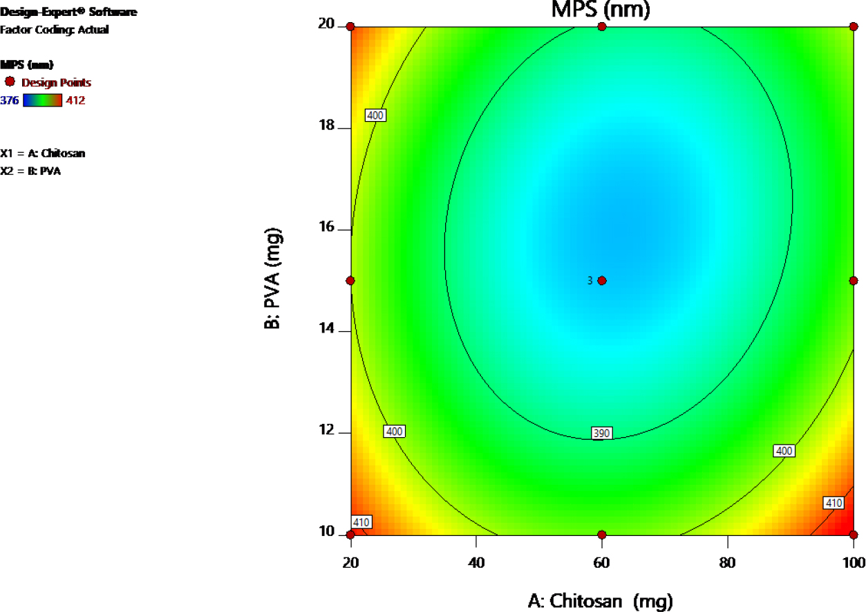
Figure 4A:
Counter plot of Y1 (Particles Size).
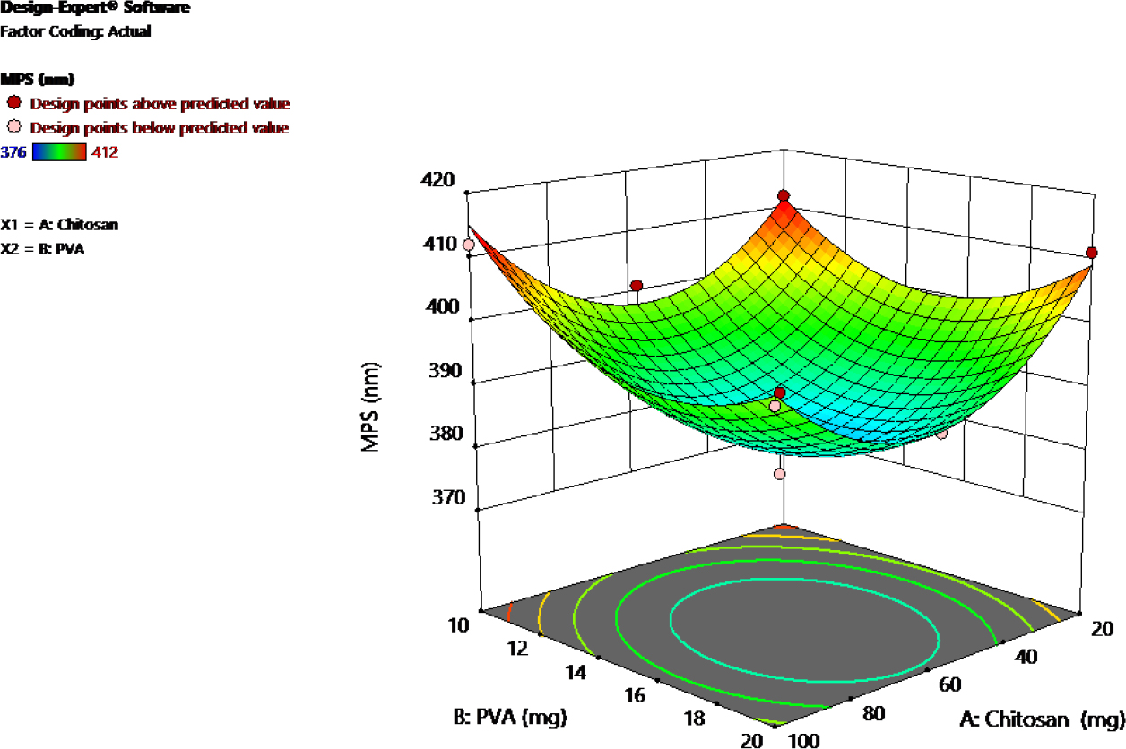
Figure 4B:
3D response Surface plot of Y1 (Particles Size).
Response 2: (particle Charge)
The nanoparticle charge, which varied between 29.87 mV and 36.76 mV as shown in Table 2, was notably affected by the concentrations of chitosan and PVA, based on the quadratic model analysis. The statistical analysis revealed an adjusted R² of 0.6795, a predicted R² of 0.2222, and a sequential p-value of 0.0401, confirming the reliability of the model. Additionally, the Lack of Fit p-value of 0.7263 supports a strong fit. The perturbation plot (Figure 5A) and the contour plots (Figure 5B) provide further insight into the relationships between the factors.
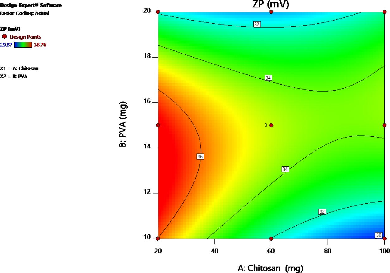
Figure 5A:
Counter plot of Y2 (Particles Charge).
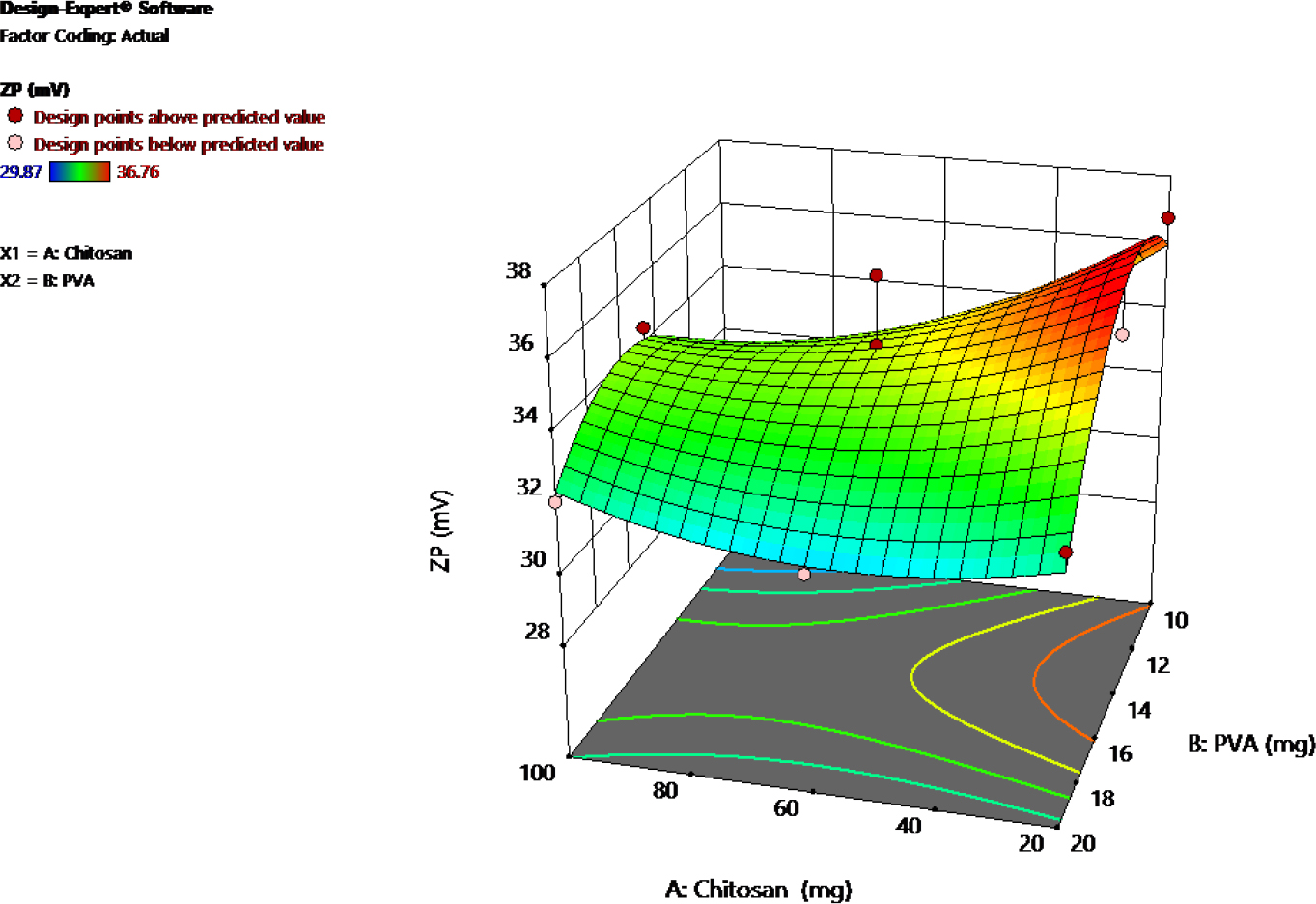
Figure 5B:
3D response Surface plot of Y2 (Particles Charge).
Optimisation by Desirability Function
The optimization process uses the desirability function to adjust parameters within specified ranges. Chitosan (20-100 mg) and Polyvinyl Alcohol (10-20 mL) are set within limits, while MPS is minimized (376-412 nm) and ZP is maintained (29.87-36.76 mV). The goal is to find the best combination of these values for optimal outcomes. After applying the constraints, optimized values for Chitosan (62.575 mg), Polyvinyl Alcohol (16.080 mL), MPS (382.700 nm), and ZP (34.310 mV) were chosen, yielding a desirability score of 0.814 in Figure 6. These values were applied to Response 1 and Response 2, resulting in observed values of MPS at 337.8 nm (Figure 7) and ZP at 32.4 mV (Figure 8).
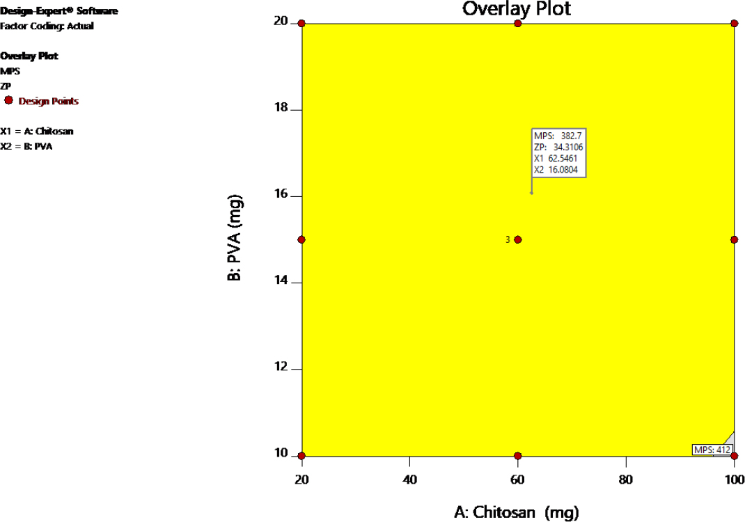
Figure 6:
Optimized desirability overlay Plot.
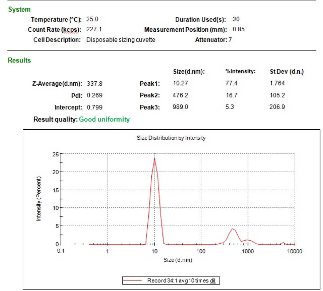
Figure 7:
Particles size of optimized batch.
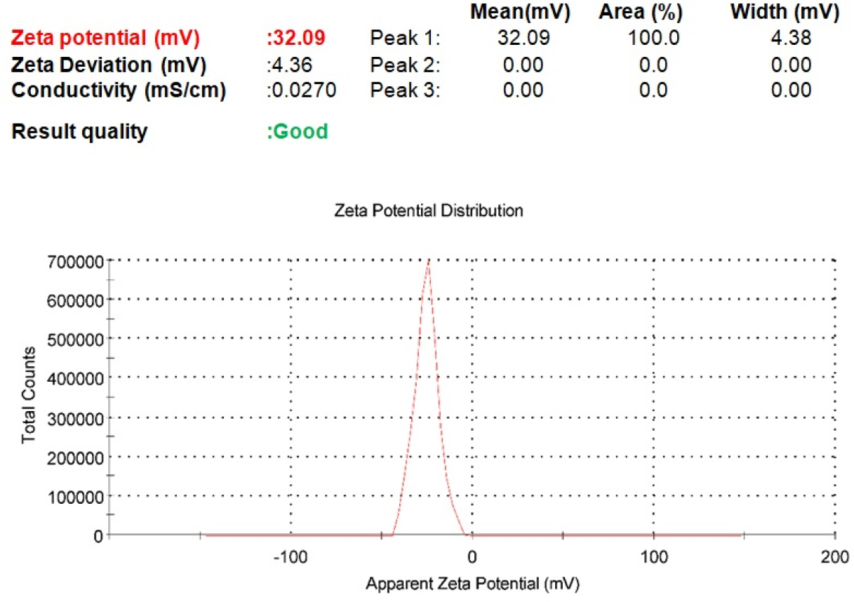
Figure 8:
Particles Charge of optimized batch.
Drug loading and Entrapment efficiency
The percentages of drug loading and entrapment efficiency of the optimised batch were 22.96±1.23% and 81.28±56%.
In vitro drug release study
The in vitro drug release profile of free drug and the palbociclib loaded polymeric nanoparticles formulation was determined by using dialysis bag method in Table 3 and Figure 9. The free drug exhibited rapid release (50% in 2 hr; 90% in 12 hr). In contrast, the nanoparticles showed a sustained profile (50% in 11.8 hr; 90% in 26.1 hr), due to the polymer matrix barrier.
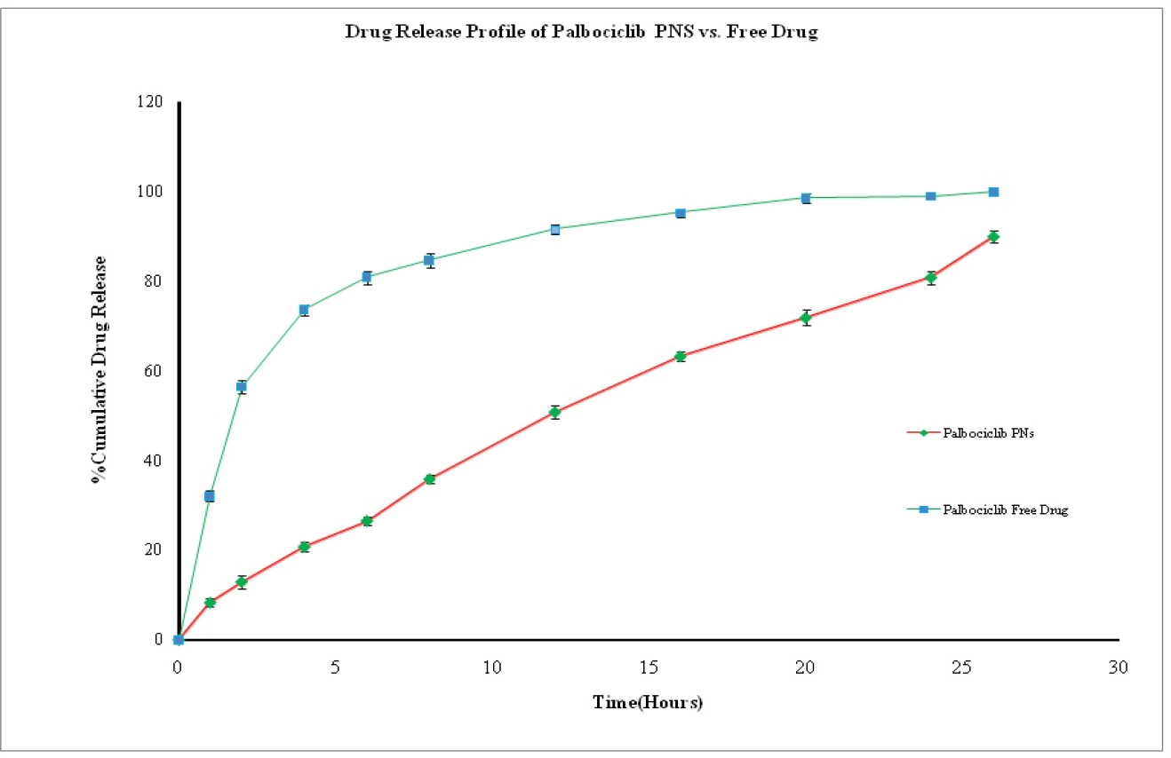
Figure 9:
Graph Plot of In vitro % Cumulative Drug Release of Optimized batch.
| % of Palbociclib PNS Drug Release | % of Free Drug Release | |||||||
|---|---|---|---|---|---|---|---|---|
| Time(hr) | R1 | R2 | R3 | Mean±SD | R1 | R2 | R3 | Mean±SD |
| 0 | 0 | 0 | 0 | 0 | 0 | 0 | 0 | 0 |
| 1 | 8.41 | 7.27 | 9.28 | 8.32±1.01 | 30.78 | 32.18 | 33.13 | 32.03±1.18 |
| 2 | 14.55 | 12.62 | 11.63 | 12.93±1.49 | 54.87 | 56.69 | 57.74 | 56.43±1.45 |
| 4 | 20.66 | 21.81 | 19.73 | 20.73±1.04 | 72.15 | 74.28 | 74.23 | 73.55±1.22 |
| 6 | 25.61 | 26.33 | 27.64 | 26.53±1.03 | 82.41 | 80.55 | 79.47 | 80.81±1.49 |
| 8 | 34.81 | 35.89 | 36.85 | 35.85±1.02 | 86.21 | 84.36 | 83.25 | 84.61±1.50 |
| 12 | 51.05 | 52.12 | 49.13 | 50.77±1.52 | 91.51 | 92.52 | 90.53 | 91.52±1.00 |
| 16 | 63.79 | 61.92 | 63.84 | 63.18±1.09 | 94.01 | 95.41 | 96.02 | 95.15±1.03 |
| 20 | 73.78 | 70.89 | 70.75 | 71.81±1.71 | 97.43 | 98.46 | 99.45 | 98.45±1.01 |
| 24 | 81.07 | 79.14 | 82.12 | 80.78±1.51 | 98.59 | 98.62 | 99.21 | 98.81±0.35 |
| 26 | 88.28 | 90.42 | 90.97 | 89.89±1.42 | 99.79 | 99.81 | 99.84 | 99.81±0.03 |
In vitro drug release kinetics model
The in vitro drug release kinetics of the optimized formulation of Palbociclib PNS was evaluated using the “DD Solver” software shows in Table 4; Korsmeyer-Peppas model (R2=0.997) and release exponent (n=0.762) suggests anomalous (non-fickian) transport.
| Optimized Batch | Zero Order (r2) | First order (r2) | Higuchi (r2) | Hixson-Crowel (r2) | Koysmeyer-Peppas (r2) |
| 0.9689 | 0.9848 | 0.9391 | 0.9944 | 0.9977 |
Morphological Characterisation of optimised batch
The surface morphology of the optimized Palbociclib-loaded nanoparticles was examined using Scanning Electron Microscopy (SEM). The images (Figure 10) revealed uniform, spherical nanoparticles with smooth surfaces and no evidence of aggregation, confirming the homogeneity of the optimized formulation.

Figure 10:
Image of SEM of the optimized batch.
DISCUSSION
Palbociclib-loaded oral Polymeric Nanoparticles (PNS) were developed using a solvent evaporation method, with formulation optimization guided by a two-factor Central Composite Design (CCD) in Design Expert® software version 11 (Version 7.0, Stat Ease Inc., USA). The independent variables chitosan (100-200 mg) and PVA (10-20 mg) were systematically varied to analyze their effects on critical responses including particle size and zeta potential. Through response surface methodology and overlay plotting, the software facilitated the identification of an optimized nanoparticle formulation with desired characteristics (Sandhya, 2022).
FT-IR analysis showed minor peak shifts: C=O (1600.59→1601.79 cm⁻¹), N-H (3699.56→3506.30 cm⁻¹), and C≡N (2947.63→2922.80 cm⁻¹). These indicate physical interactions like hydrogen bonding but confirm no chemical incompatibility, ensuring formulation stability (Sandhya, 2022).
The nanoparticle size, ranging from 376 to 412 nm, was predominantly influenced by chitosan concentration. Statistical analysis confirmed a robust model fit (adjusted R²: 0.8150; p-value: 0.0035), indicating high reliability. Particle charge, varying between 29.87 and 36.76 mV, was significantly affected by both chitosan and PVA concentrations, with the model also demonstrating a reliable fit (adjusted R²: 0.6795; p-value: 0.0401). These results validate the quadratic model’s effectiveness in optimizing both critical quality attributes (Xieet al., 2008, Yanget al., 2010).
The desirability function was employed for optimization, with chitosan (20-100 mg) and PVA (10-20 mL) as variables. Targets were to minimize particle size (376-412 nm) and maintain zeta potential (29.87-36.76 mV). The optimal formulation used 62.58 mg chitosan and 16.08 mL PVA, achieving a high desirability of 0.814. Experimental validation confirmed a particle size of 337.8 nm and zeta potential of 32.4 mV, aligning well with predicted outcomes (Sandhya, 2022, Liuet al., 2010).
An in vitro release study compared free palbociclib to polymeric nanoparticles using a dialysis method. The free drug displayed rapid release (50% in 2 hr; 90% in 12 hr), while the nanoparticles demonstrated sustained release (50% in 11.8 hr; 90% in 26.1 hr), attributed to the polymer matrix barrier (Yuanet al., 2012). DD Solver analysis indicated the release kinetics followed the Korsmeyer-Peppas model (R²=0.997), with an exponent (n=0.762) confirming anomalous (non-Fickian) diffusion-erosion release mechanisms (Yuanet al., 2012, Chakrabortyet al., 2025).
The optimized batch exhibited a drug loading of 22.96%±1.23 and an entrapment efficiency of 81.28% ±0.56, confirming efficient nanoparticle drug incorporation (Xieet al., 2008).
The optimized palbociclib-loaded nanoparticles displayed a smooth, spherical morphology under SEM examination. This uniform structural characteristic supports predictable drug release profiles, enhances formulation stability, and promotes efficient cellular uptake, collectively validating the effectiveness of the preparation technique (Fanget al., 2011).
CONCLUSION
Palbociclib-loaded nanoparticles were successfully developed. FT-IR analysis confirmed excipient compatibility without chemical interactions. Chitosan and PVA concentrations effectively controlled nanoparticle characteristics, yielding a size of 337.8 nm and zeta potential of 32.4 mV, which aligned closely with predicted values. The formulation exhibited sustained drug release via diffusion-erosion mechanisms, high drug loading (22.96%), excellent entrapment efficiency (81.28%), and uniform spherical morphology, collectively demonstrating its suitability as an effective oral drug delivery system.
Cite this article:
Hoque N, Choudhury A, Datta D. Formulation, Central Composite Design Optimization and in vitro Evaluation of Folic Acid Conjugated Palbociclib Loaded Polymeric Nanoparticles. J Young Pharm. 2025;17(4):877-86.
ACKNOWLEDGEMENT
The authors thank Assam Down Town University for its resources and the faculty and staff of Pharmaceutical Science for their advice and technical contributions.
ABBREVIATIONS
| CCD | Central Composite Design |
|---|---|
| PVA | Polyvinyl Alcohol |
| PNS | Polymeric Nanoparticles |
| SEM | Scanning Electron Microscope |
| CDK | Cyclin-Dependent Kinase |
References
- Aldaais E.. (2018) Generalized theory of monoligand-receptor binding for the improvement of nanoparticle design.. Journal of Nanomedicine and Nanotechnology 09 https://doi.org/10.4172/2157-7439.1000512 | Google Scholar
- Amanzio M., Vase L., Mitsikostas D. D.. (2020) [Editorial]. Editorial: Nocebo effects and their influence on clinical trials and practice: Modulating factors in healthy and pathological conditions.. Frontiers in Pharmacology 11: 100 https://doi.org/10.3389/fphar.2020.00100 | Google Scholar
- Chakraborty P., Das A., Chatterjee S., Bairagi A., Bhattacharya H., Bhattacharyya C., Chatterjee N., Sil P. C., Dewanjee S., et al. (2025) Formulation and evaluation of polymeric nanoparticles to improve chemotherapeutic efficacy of mangiferin against breast cancer.. Naunyn-Schmiedeberg’s Archives of Pharmacology 398: 12433-12448 https://doi.org/10.1007/s00210-025-04068-0 | Google Scholar
- Fang J., Nakamura H., Maeda H.. (2011) The EPR effect: Unique features of tumor blood vessels for drug delivery, factors involved, and limitations and augmentation of the effect.. Advanced Drug Delivery Reviews 63: 136-151 https://doi.org/10.1016/j.addr.2010.04.009 | Google Scholar
- Kankala R. K., Xu P.-Y., Chen B.-Q., Wang S.-B., Chen A.-Z.. (2021) Supercritical fluid (SCF)-assisted fabrication of carrier-free drugs: An eco-friendly welcome to active pharmaceutical ingredients (APIs).. Advanced Drug Delivery Reviews 17(4)6: Article 113846 https://doi.org/10.1016/j.addr.2021.113846 | Google Scholar
- Khan I., Saeed K., Khan I.. (2019) Nanoparticles: Properties, applications and toxicities.. Arabian Journal of Chemistry 12: 908-931 https://doi.org/10.1016/j.arabjc.2017.05.011 | Google Scholar
- Krishnakumar N., Sulfikkarali N., RajendraPrasad N., Karthikeyan S.. (2011) Enhanced anticancer activity of naringenin-loaded nanoparticles in human cervical (HeLa) cancer cells.. Biomedicine and Preventive Nutrition 1: 223-231 https://doi.org/10.1016/j.bionut.2011.09.003 | Google Scholar
- Liu Y., Li K., Pan J., Liu B., Feng S.-S.. (2010) Folic acid conjugated nanoparticles of mixed lipid monolayer shell and biodegradable polymer core for targeted delivery of docetaxel.. Biomaterials 31: 330-338 https://doi.org/10.1016/j.biomaterials.2009.09.036 | Google Scholar
- Lu Z., Zhang Y., Wang Y., Tan G.-H., Huang F.-Y., Cao R., He N., Zhang L., et al. (2021) A biotin-avidin-system-based virus-mimicking nanovaccine for tumor immunotherapy.. Journal of Controlled Release 332: 245-259 https://doi.org/10.1016/j.jconrel.2021.02.029 | Google Scholar
- Mandal B., Bhattacharjee H., Mittal N., Sah H., Balabathula P., Thoma L. A., Wood G. C., et al. (2013) Core-shell-type lipid-polymer hybrid nanoparticles as a drug delivery platform.. Nanomedicine: Nanotechnology, Biology, and Medicine 9: 474-491 https://doi.org/10.1016/j.nano.2012.11.010 | Google Scholar
- Martin A.-M.. (2000) Genetic and hormonal risk factors in breast cancer.. Journal of the National Cancer Institute 92: 1126-1135 https://doi.org/10.1093/jnci/92.14.1126 | Google Scholar
- Patra D. C. N.. (2018) Solid lipid-based delivery system for oral delivery of drugs: A review.. Asian Journal of Pharmaceutics 12: 345-354 https://doi.org/10.22377/ajp.v12i04.2902 | Google Scholar
- Rocca A., Schirone A., Maltoni R., Bravaccini S., Cecconetto L., Farolfi A., Bronte G., Andreis D., et al. (2017) Progress with palbociclib in breast cancer: Latest evidence and clinical considerations.. Therapeutic Advances in Medical Oncology 9: 83-105 https://doi.org/10.1177/1758834016677961 | Google Scholar
- Sandhya B. H. K.. (2022) Palbociclib loaded solid lipid nanoparticles by box-Behnken design: Solubility enhancement.. International Journal of Pharmaceutical Sciences and Research 13: 1241-1250 https://doi.org/10.13040/IJPSR.0975-8232.13(3).1241-50 | Google Scholar
- Sartaj A., Qamar Z., Qizilbash F. F., Annu S., Alhakamy N. A., Baboota S., Ali J., et al. (2021) Polymeric nanoparticles: Exploring the current drug development and therapeutic insight of breast cancer treatment and recommendations.. Polymers 13: Article 4400 https://doi.org/10.3390/polym13244400 | Google Scholar
- Schaffazick S. R., Pohlmann A. R., Dalla-Costa T., Guterres S. S.. (2003) Freeze-drying polymeric colloidal suspensions: Nanocapsules, nanospheres and nanodispersion. A comparative study.. European Journal of Pharmaceutics and Biopharmaceutics 56: 501-505 https://doi.org/10.1016/S0939-6411(03)00139-5 | Google Scholar
- Xie S., Wang S., Zhao B., Han C., Wang M., Zhou W., et al. (2008) Effect of PLGA as a polymeric emulsifier on preparation of hydrophilic protein-loaded solid lipid nanoparticles.. Colloids and Surfaces. B, Biointerfaces 67: 199-204 https://doi.org/10.1016/j.colsurfb.2008.08.018 | Google Scholar
- Yang C.-R., Zhao X.-L., Hu H.-Y., Li K.-X., Sun X., Li L., Chen D.-W., et al. (2010) Preparation, optimization and characteristic of huperzine A loaded nanostructured lipid carriers.. Chemical and Pharmaceutical Bulletin 58: 656-661 https://doi.org/10.1248/cpb.58.656 | Google Scholar
- Yu K., Zhao J., Yu C., Sun F., Liu Y., Zhang Y., Lee R. J., Teng L., Li Y., et al. (2016) Role of four different kinds of Polyethylenimines (PEIs) [In preparation] of Polymeric Lipid Nanoparticles and Their Anticancer Activity Study.. Journal of Cancer 7: 872-882 https://doi.org/10.7150/jca.13855 | Google Scholar
- Yuan Y.-Y., Mao C.-Q., Du X.-J., Du J.-Z., Wang F., Wang J., et al. (2012) Surface charge switchable nanoparticles based on zwitterionic polymer for enhanced drug delivery to tumor.. Advanced Materials 24: 5476-5480 https://doi.org/10.1002/adma.201202296 | Google Scholar
