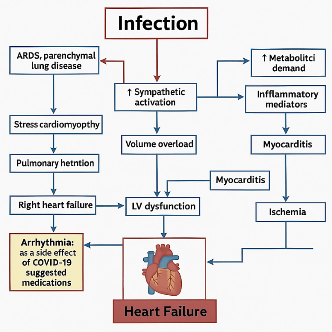
ABSTRACT
ABSTRACT
There is growing evidence highlighting a significant risk of cardiovascular complications in patients recovering from COVID-19, particularly among those experiencing long COVID or persistent post-acute symptoms. Numerous studies have shown that COVID-19 can trigger or exacerbate cardiovascular conditions such as myocarditis, arrhythmias, thromboembolic events, and heart failure, even in individuals without a prior history of cardiovascular disease. The objective of this review is to evaluate the prevalence and risk of cardiovascular complications following COVID-19 infection. By synthesizing current research findings, this paper aims to provide a comprehensive understanding of how COVID-19, especially in its long-term manifestations, contributes to increased cardiovascular risk. A systematic literature search was conducted using PubMed, Scopus, and Google Scholar to identify relevant studies. Key findings indicate an increase in cardiovascular mortality across countries during 2020, with COVID-19 significantly contributing to the incidence of myocarditis, arrhythmias, heart failure, and thrombotic events-particularly among older adults and those with pre-existing conditions. Long COVID is often associated with persistent cardiac manifestations, driven by inflammation, endothelial dysfunction, and hypercoagulability. The findings highlight the importance of managing chronic conditions and establishing specialized clinics for ongoing care. Routine cardiovascular screening, comorbidity management, and vaccination are essential components in preventing and mitigating cardiovascular complications in post-COVID patients.
INTRODUCTION
The global impact of COVID-19 has been substantial, with approximately 704.8 million confirmed cases and 7.01 million deaths reported (Harriset al., 2021, and Lancet, 2021). Severe COVID-19 symptoms are experienced by a small percentage of patients whose illness includes coagulopathy, inflammation, and microvascular damage. This condition can result in myocardial damage, thromboembolism, and occlusive events in the arteries. Individuals who already have cardiovascular disease or have risk factors for it may be more vulnerable. Patients frequently arrive with fever, cough, dyspnea, myalgia, and exhaustion; sputum production, hemoptysis, headache, and diarrhea are less frequent. Respiratory syndrome coronavirus has been linked to myocardial injury (Pellicoriet al., 2022) and myocarditis with elevated troponin. These conditions are believed to be caused by direct myocardial injury, increased cardiac physiologic stress, or hypoxia. A study of 41 patients with COVID-19 in Wuhan, China, revealed that 5 patients (12%) had a high sensitivity troponin above the threshold of 28 pg/mL. This was one of the first reports of myocardial injury linked to SARS-CoV-2. According to later research, 7-17% of patients hospitalized with COVID-19 and 23%-31% of patients admitted to the ICU may experience myocardial injury with elevated troponin levels. The risk of AMI and disruption of atherosclerotic plaque is increased by severe systemic inflammation. A 2018 study discovered that, within the first seven days of a disease diagnosis, influenza and a few other viral illnesses were linked to a higher risk of Acute Myocardial Infarction (AMI), with an incidence ratio of 6.1 for influenza and 2.8 for other viruses. The American College of Cardiology states that while fibrinolysis may be considered in those with “low-risk STEMI,” defined by inferior STEMI with no right ventricular involvement or lateral AMI with hemodynamic compromise, percutaneous coronary intervention is more commonly performed at most institutions and remains the treatment of choice (Xieet al., 2022).
This literature review seeks to discuss the late cardiovascular sequelae and risks in individuals who have survived COVID-19. This is mainly aimed at determining how vast the role is of COVID-19 in causing cardiovascular dysfunction, specific to either direct viral pathophysiological changes or indirectly due to related comorbidities and lifestyle considerations. This review provides a comprehensive insight into the link between SARS-CoV-2 infection and cardiovascular health by examining clinical outcomes in post-COVID-19 patients and their association with the onset or worsening of Cardiovascular Diseases (CVDs). It explores the underlying pathophysiological mechanisms responsible for cardiovascular complications in the post-COVID setting, such as systemic inflammation, endothelial dysfunction, and virus-induced coagulopathies. The review also analyzes the prevalence and nature of circulatory disorders in recovered patients, while evaluating the role of pre-existing conditions and lifestyle factors in elevating cardiovascular risk. Furthermore, it considers whether these cardiovascular manifestations should be regarded as direct consequences of viral infection or as secondary outcomes of the body’s inflammatory response. Ultimately, the review highlights growing evidence of the increased cardiovascular burden linked to COVID-19 and underscores the importance of long-term cardiovascular monitoring and preventive strategies for effectively managing post-COVID-19 patients.
METHODOLOGY
A wide survey of scientific literature provided with various databases, such as Scopus, Web of Science, and Google Scholar, has been implemented to carry out this review.
Global Infection Estimates
Actual infection rates are likely higher due to underreporting and limited testing. The image provides comprehensive global and country-specific data on COVID-19 cases and trends. As of April 2025, the number of COVID-19 cases reported to the WHO was Total cumulative reported cases: 778 million. Cases reported every 28 days: 33,400 globally (Figure 1) (IMH, SPHA, CDC, ICMR, and Brazilian Ministry of Health 2020 and 2021). The COVID-19 pandemic has had a lasting impact on global health, from the immediate devastation caused by the virus itself to the longer-term consequences like long COVID, mental health crises, and exacerbated health inequities (Figure 2). A large meta-analysis of nearly 10 million people found significantly higher cardiovascular risks in those with long COVID. Myocarditis risk was 6.11 times higher, followed by increased risks for cardiogenic shock (2.09x), heart failure (1.72x), stroke (1.71x), cardiomyopathy (1.71x), hypertension (1.70x), coronary heart disease (1.61x), and arrhythmia (1.60x) (Xieet al., 2022; Libby Pet al., 2020; Nishiga Met al., 2020). These risks are linked to endothelial damage, persistent inflammation, autoimmune responses, and disruption of the renin-angiotensin system (Figure 3).
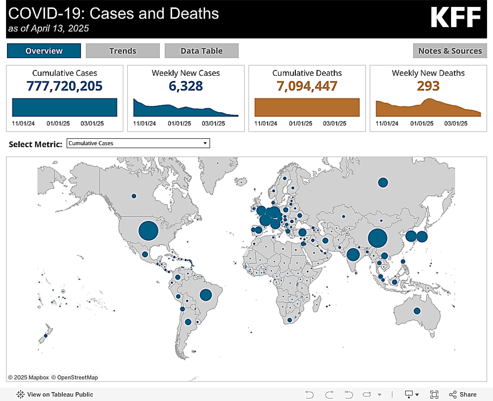
Figure 1:
Number of COVID-19 cases reported to WHO (cumulative total) Global health Consequences of post COVID.
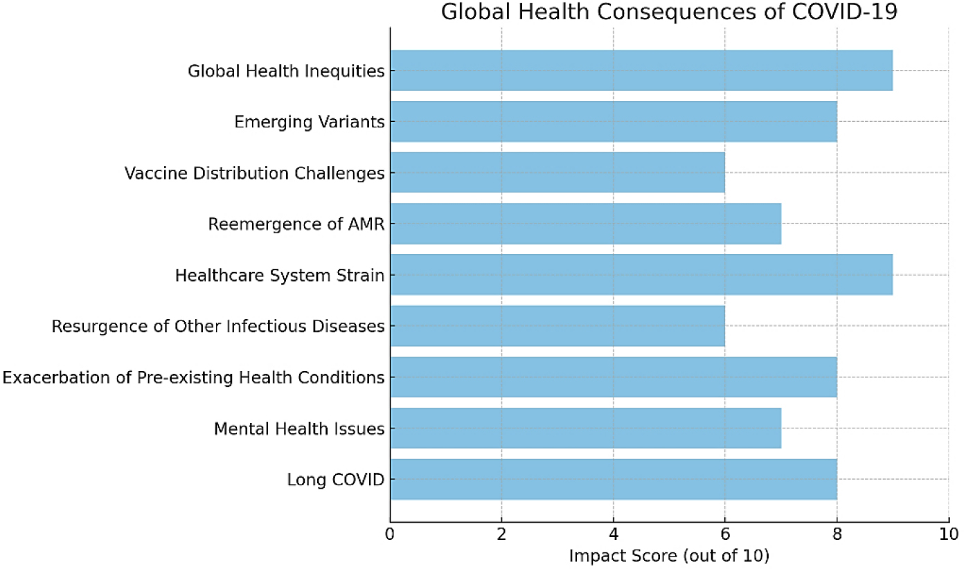
Figure 2:
The relative impact of various post-COVID health consequences on a scale of 1 to 10.
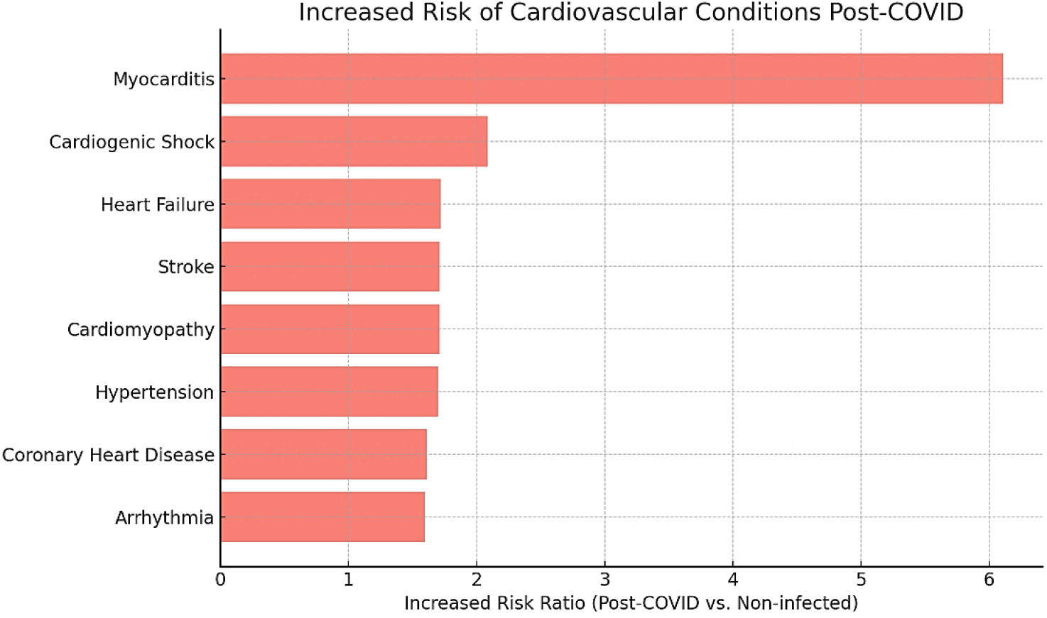
Figure 3:
The increased risk of various cardiovascular conditions in individuals post-COVID.
In 2020, excess cardiovascular deaths per 1 million population were notably high in certain countries, with Czechia reporting the highest globally at 983 deaths per 1 million, followed by India with 243 deaths per 1 million and Sri Lanka with 238 deaths per 1 million. Age-wise patterns of excess mortality revealed that individuals aged over 60 years experienced the most significant increase in deaths across all countries studied. In the middle-aged group (40-59 years), moderate excess deaths were observed, particularly in countries like Nigeria, Argentina, and the USA. Conversely, young adults (20-39 years) and children (0-19 years) experienced minimal or no excess deaths. Country-specific trends showed that the Czech Republic, the USA, and Spain had among the highest excess mortality in the elderly, while Australia, Japan, and Switzerland exhibited lower excess mortality across all age groups. An unusual pattern was noted in Nigeria, where there was a significant elevation in excess deaths in the middle-aged group, suggesting potential healthcare disparities in that population (Furuse 2023) (Figure 4).
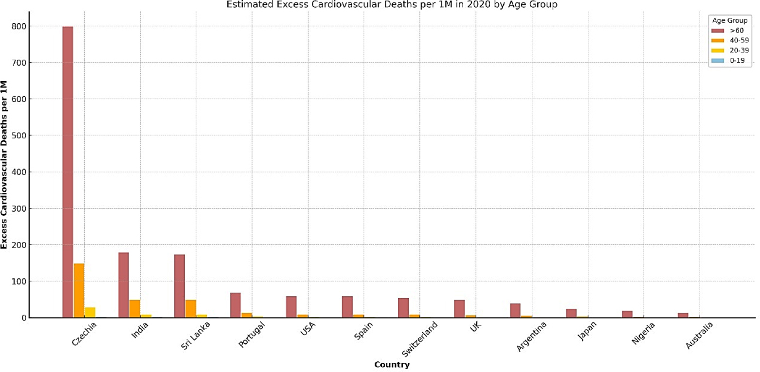
Figure 4:
Estimated Excess Cardiovascular Deaths per 1 Million Population in 2020 (Based on seroprevalence data).
The study by Gottlieb et al., (2020) offers a comprehensive overview of cardiovascular complications and thrombotic risks in COVID-19 patients, emphasizing the significant burden of these conditions in both hospitalized and critically ill individuals. Cardiac complications were notably prevalent, with cardiomyopathy reported in 33% of cases, acute heart failure in 23%, dysrhythmias in 17% of hospitalized patients and 44% of those in the ICU, and palpitations occurring in 7%. In terms of thrombotic events, the study highlights Deep Vein Thrombosis (DVT) in 21% of patients and Pulmonary Embolism (PE) in 32%, with ICU patients showing a markedly higher incidence of PE at 65%. The analysis further demonstrates a strong correlation between elevated D-dimer levels and thrombotic risk, with median D-dimer values significantly higher in patients with PE (11.07 µg/mL) compared to those with general COVID-19 pneumonia (6.06 µg/mL). Moreover, D-dimer levels exceeding 1 µg/mL were associated with an 18.4-fold increase in the risk of in-hospital mortality, underscoring the prognostic value of this biomarker in COVID-19-related cardiovascular complications (Figure 5).
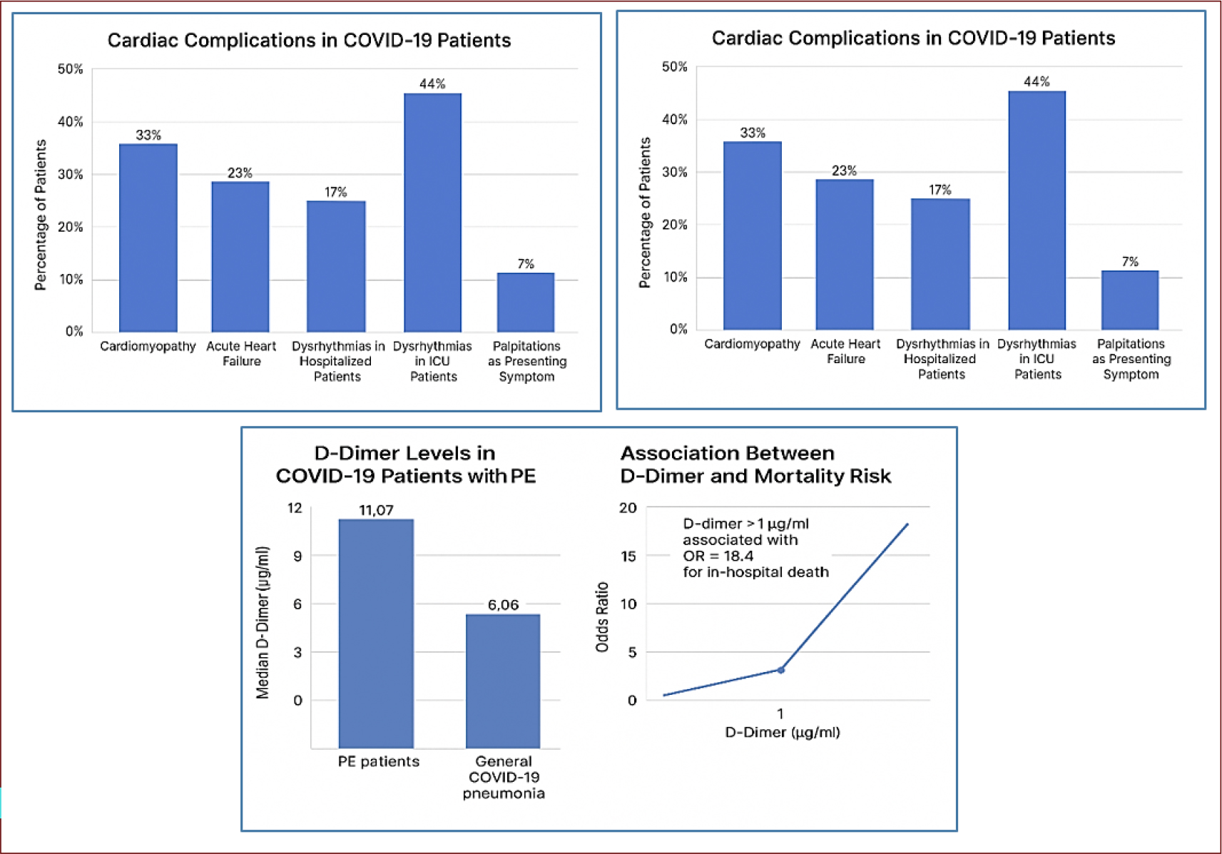
Figure 5:
A comprehensive view of cardiovascular complications and thrombotic risks in COVID-19 patients. (Gottlieb Met al., 2020).
Pathophysiology of Post-COVID Cardiovascular Disease: Mechanisms Linking COVID-19 to Cardiovascular Disease (CVD)
Endothelial dysfunction and vascular inflammation
Since the outbreak of COVID-19 in early 2020, emerging evidence has demonstrated endothelial dysfunction as the unifying and central mechanism operating to collectively indicate the alterations in vascular homeostasis in COVID-19 by overviewing the most recent literature as summarized in Table 1 and Figure 6 (Xuet al., 2023, and Mukkawaret al., 2024).
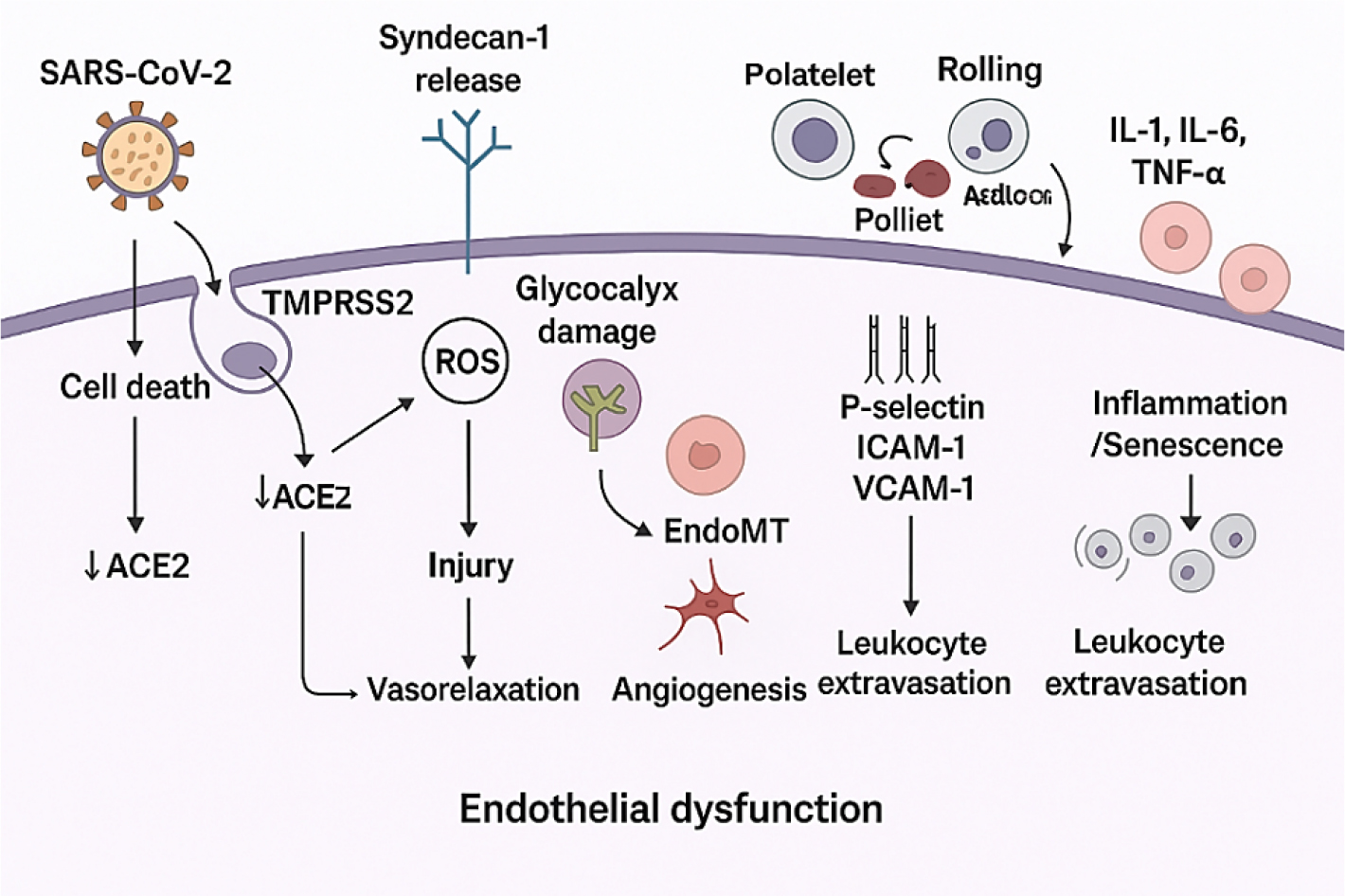
Figure 6:
Processes culminate in endothelial dysfunction, characterized by inflammation, coagulation abnormalities, increased permeability, and organ damage.
| Key steps | Pathways |
|---|---|
| Viral Entry and Binding | – SARS-CoV-2 binds to ACE2 and Neuropilin-1 on endothelial cells. – TMPRSS2 primes the spike protein, enabling entry. – Leads to ACE2 downregulation and cell death. |
| Glycocalyx Damage | – Infection induces syndecan-1 release. – Causes degradation of the glycocalyx layer, compromising vascular integrity. |
| Reactive Oxygen Species (ROS) | – Viral infection elevates ROS production. – Results in cellular injury, nitric oxide (NO) depletion, and impaired vasorelaxation. |
| EndoMT (Endothelial-to-Mesenchymal Transition) | – Induced by TNF-α, IL-1β, and TGF-β1. – Leads to endothelial dysfunction and fibrosis. |
| Angiogenesis | – Vascular endothelial growth factor (VEGF) promotes new blood vessel formation. – May be compensatory or pathological. |
| Leukocyte and Platelet Activation | – Endothelial cells express adhesion molecules (P-selectin, ICAM-1, VCAM-1). – Promotes leukocyte rolling, adhesion, and extravasation. |
| Cytokine Storm and Inflammation | – Elevated IL-1, IL-6, TNF-α levels. – Causes systemic inflammation, senescence, and cytokine storm. |
Hypercoagulability and thrombotic risks
Much literature reports that the changes in the coagulation system of COVID-19 patients generally show enhanced coagulation and thrombosis. Complications of severe and critical COVID-19 include ARDS, embolism or pulmonary thrombosis, and hypercoagulable state. COVID-19 is accompanied by a hypercoagulable state, and the occurrence of thrombotic diseases and microthrombi can be seen in cardiac vessels, the hepatic portal area, and renal interstitium; the autopsy pathology of COVID-19 confirmed this. COVID-19-related hypercoagulant and thrombotic complications may be due to the following pathological mechanisms.
There are 2 aspects of factors for the formation of hypercoagulability and thrombotic disease: one is the enhancement of coagulation, and the other is the weakening of anticoagulation and fibrinolysis. From the coagulation cascade, we can see that the enhancement of coagulation can be initiated through an intrinsic or extrinsic pathway, causing the chain-level expansion of coagulation factors and thrombin formation. Many factors are activators of the coagulation system, and damage to the vascular endothelium and extrinsic tissue factors are the main promoters of coagulation. The core of the fibrinogen and fibrin monomer cross-linking. Plasmin is activated by plasminogen under the action of t-PA and then quickly combines with 2-antiplasmin to form the Plasmin-Antiplasmin (PAP) complex. Vascular endothelial cells secrete t-PA, which is usually in the form of a tPA-PAI-1 Complex (tPAIC), and there is abundant Thrombomodulin (TM) under vascular endothelial cells, which can be activated by thrombin, thereby activating the protein C pathway anticoagulants. Therefore, PAP, TPAIC, and TM can be used as molecular markers for early monitoring of hypercoagulability and micro thrombosis in blood vessels. Vascular endothelial cells are an important link in regulating the coagulation, anticoagulation, and fibrinolysis systems and are closely related to the body’s hypercoagulable state and the occurrence of thrombotic diseases. This may lead to myocardial dysfunction and damage, endothelial dysfunction, microvascular dysfunction, plaque instability, and Myocardial Infarction (MI). (Valencia Iet al., 2024)
SARS-CoV-2 interaction with ACE2 disrupts the Renin-Angiotensin System (RAS), contributing to lung injury or protection depending on the signaling balance (Table 2).
| Key Process | Mechanism |
|---|---|
| Viral Entry and Replication | SARS-CoV-2 binds to ACE2 via the S-protein, primed by TMPRSS2. – Virus enters the host cell and replicates, causing ACE2 downregulation. |
| ACE2 Downregulation | – Reduces the conversion of Ang II to Ang 1-7, disrupting the protective RAS axis. |
| Angiotensin Pathways | ACE converts Angiotensin I to Ang II, which acts on AT₁R, causing: Pro-oxidation, Inflammation, Vasoconstriction, Fibrosis – Leads to lung injury. |
| Protective RAS Axis (ACE2 Pathway) | ACE2 converts Angiotensin II to Ang 1-7, activating MasR and AT₂R receptors. – Results in: Anti-inflammation, Vasodilation, Anti-fibrosis – Provides lung protection. |
| ADAM17 Role | Induced by viral infection, ADAM17 promotes ACE2 shedding, further decreasing ACE2 activity. |
| rhACE2 (Recombinant Human ACE2) | Proposed as a therapeutic agent to bind the virus and protect native ACE2, potentially preserving RAS balance. |
Myocardial injury and fibrosis
Table 3 integrates both direct viral effects and host immune/inflammatory responses as contributors to cardiac injury in COVID-19 (Babapoor-Farrokhran S et al., 2024).
| Key Mechanism | Pathways |
|---|---|
| ACE2 Receptor Disruption | SARS-CoV-2 binds to ACE2 on cardiomyocytes, leading to ACE2 downregulation. This impairs Angiotensin 1-7 signaling, contributing to cardiac dysfunction. |
| Direct Viral Invasion of Myocytes | SARS-CoV-2 RNA has been detected in autopsied heart tissues, suggesting direct viral invasion and damage to heart muscle cells. |
| Myocardial Inflammation | Infiltration of myocardial macrophages and inflammation observed in post-mortem samples, indicating immune-mediated damage. |
| Increased TNF-α Production | ACE2 downregulation increases TNF-α, a pro-inflammatory cytokine associated with cardiac injury. |
| TGF-β/Smad Signaling Pathway Activation | Known to be activated by SARS-CoV and involved in lung and myocardial fibrosis; a potential mechanism of cardiac remodeling. |
| Systemic Inflammation (Cytokine Storm) | Exaggerated immune response with elevated cytokines (e.g., TNF-α, IL-6, CRP) causing cardiomyocyte damage, especially in patients with preexisting CVD. |
| Elevated Troponin T (TnT) and CRP | Clinical biomarker evidence of myocardial injury linked to inflammation in severe COVID-19 cases. |
| Type 1 and Type 2 Helper T Cell Imbalance | Suggested to contribute to transition from innate to adaptive immunity, possibly overreacting and contributing to cardiac damage. |
| Exaggerated Interferon-Mediated Response | Strong interferon responses may damage myocardial tissue in severe SARS and COVID-19 infections. |
| Myocardial Interstitial Fibrosis | Fibrotic remodeling due to inflammation and cytokine signaling, contributing to long-term cardiac dysfunction. |
| Hypoxia-Induced Injury | Respiratory failure in COVID-19 can reduce oxygen supply, leading to myocardial ischemia and injury. |
| Coronary Plaque Destabilization | Inflammatory response may destabilize atherosclerotic plaques, leading to acute coronary events such as myocardial infarction. |
Immune-mediated mechanisms
Pathogenic agents can infect cells, triggering humoral and cellular immune responses in the host that are necessary to eradicate the viral infection. On the other hand, an unchecked or inadequate immune response can result in immunopathology and seriously harm patients. While lowering the possible risk of inflammation, a better understanding of the immune response brought on by SARS-CoV-2 infection may result in the development of novel immunotherapies. Like SARS-CoV, the cell surface receptor ACE2 mediates the most well-known cellular infection mechanism of SARS-CoV-2. Numerous investigations have demonstrated that SARS-CoV-2 could infect cells that were genetically altered to express only the ACE2 receptor. Since the human lung and small intestine epithelia primarily contain this ACE2 receptor, SARS-CoV-2 is more likely to infect the respiratory and gastrointestinal systems. Hikmet et al., posted the preprint, which shows that the seminal vesicle’s glandular cells, renal proximal tubules, cardiomyocytes, testicular Sertoli cells, Leydig cells, and gallbladder epithelium all expressed ACEE2.
Furthermore, it has been proposed that the virus may also infect the brain because COVID-19 patients exhibit neurological symptoms, including hyposmia in the early stages of infection, as well as headache, nausea, vomiting, and cerebral damage in more severe cases. It has been proposed that SARS-CoV-2 enters the brain through the cerebral circulation, the cribriform plate near the olfactory bulb, or the ACE2 receptor on the endothelium. These three possible routes could account for at least some of the altered sense of smell. Glial cells and, in fact, neurons are known to express ACE2, being, therefore, potential targets for SARS-CoV-2 (Babapoor-Farrokhran S et al., 2024).
Transmembrane Protease Serine 2 (TMPRSS2)
Angiotensin-Converting Enzyme 2 (ACE2). L-SIGN (CD209L) is a possible SARS-CoV-2 receptor that has received less attention. This type II transmembrane glycoprotein, which belongs to the C-type lectin family, is primarily expressed by endothelial and type II human lung alveolar epithelial cells. It was demonstrated that this receptor, albeit to a lesser degree than the ACE2 receptor, facilitated SARS-CoV entry into host cells. As a result, CD209L probably mediates SARS-CoV-2 entry as well.
Cardiovascular Manifestations in Post-COVID Patients
Acute and chronic cardiovascular conditions
The COVID-19 has emerged as a public health concern that is causing social and economic issues in nearly every nation on the planet. The rapid spread of the pandemic and high rates of morbidity in certain populations, which even reached high mortality rates in developed nations like the United States have put a strain on health systems. A. in which COVID-19 ranked as the third most common cause of death in 2020.
The coronavirus-2 that causes SARS-CoV-2 is the cause of COVID-19. Systemic severe inflammatory response and acute ARDS were the primary pathophysiologic mechanisms of this virus. Patients with comorbidities, such as prior cardiovascular disease, were identified early in the pandemic as having a higher risk of death and a worse clinical outcome. As the pandemic progressed, multiple reports alerted people to the possible cardiac and multisystemic involvement of COVID-19 (Arevalos V et al., 2021).
Myocarditis and pericarditis
The global COVID-19 pandemic is brought on by SARS-CoV-2. It results in symptoms that are both pulmonary and extrapulmonary. The most common and severe of the extrapulmonary symptoms is cardiac involvement. Cardiovascular symptoms of COVID-19 include myocarditis and pericarditis, which can develop without pulmonary involvement. Case presentation on Table 4 (Shah JZet al., 2020).
| Case | Age | Symptoms | Vital Signs | Physical Examination | Additional Notes |
|---|---|---|---|---|---|
| Case 1 | 19-year-old male | Fever, coughing, shortness of breath, generalized weakness (7 days duration) | Temperature: 38ºC, BP: 76/44 mmHg, Heart Rate: 126 bpm, Respiratory Rate: 45 bpm, Oxygen Saturation: 92% on room air. | Extremities warm, no edema, no heart failure signs, no murmurs, reduced air entry on both sides, normal abdominal exam | Respiratory distress, hypotension, tachypnea, tachycardia, no recent travel or known contact with ill individuals, no comorbid conditions. |
| Case 2 | 42-year-old male | Pleuritic chest pain, fever, coughing, dyspnea (1-week duration) | Temperature: 37ºC, BP: 113/74 mmHg, Heart Rate: 102 bpm, Respiratory Rate: 19 bpm, Oxygen Saturation: 99% on room air. | Distant heart sounds, normal chest examination | Appeared at ease, no recent travel or illness contacts. |
| Case 3 | 38-year-old diabetic male | History of confirmed moderate COVID-19 pneumonia, recovered (3 weeks), presenting with unusual pricking chest pain | – | – | Reported chest pain unrelated to posture, breathing, or physical activity; recovered from COVID-19 pneumonia, previously treated with antibiotics and antivirals. |
Post COVID Syndrome
Arrhythmias and palpitations
The impact of the coronavirus pandemic on humanity is undoubtedly one of the worst catastrophes in recent decades. As the pandemic progressed, there was increasing evidence that some patients developed persistent symptoms, often affecting multiple organ systems beyond the acute COVID-19 infection. In most cases, the infection causes mild cold symptoms such as fever, cough, and tiredness. However, a significant proportion of patients develop severe symptoms leading to pulmonary injury and ADRS, as well as multiple organ failure with a lethal outcome. Persistent fatigue, a change in or loss of smell, palpitations, and chest pain are common symptoms that patients complain about weeks or even months after having experienced an acute COVID-19 infection (Huseynov Aet al., 2023).
Heart failure and cardiomyopathy
Hypertension and Vascular complications
Long COVID can lead to hypertension and vascular complications through several interrelated mechanisms. The virus causes endothelial dysfunction by infecting cells via ACE2 receptors, leading to vascular inflammation and stiffness. It also disrupts the Renin-Angiotensin-Aldosterone System (RAAS), increasing angiotensin II levels, which promotes vasoconstriction and fluid retention. Persistent inflammation and oxidative stress further impair vascular function and nitric oxide signaling. Additionally, autonomic nervous system dysregulation (such as postural orthostatic tachycardia syndrome) can cause erratic blood pressure control. Long COVID is also linked to increased thrombotic activity and metabolic disturbances, all of which contribute to the development or worsening of hypertension and vascular disease (Farshidfaret al., 2021 and Marqueset al., 2023).
Thrombosis and embolism risks
Some noteworthy findings regarding COVID-19 were noted early in the pandemic: (1) a significant number of COVID-19 patients exhibit significantly abnormal coagulation parameters, especially D-dimer elevation, which is associated with mortality; and (2) a notably high incidence of thrombotic events is observed in COVID-19 patients, especially those in the intensive care unit. (3) Despite the use of prophylactic anticoagulation, a large incidence of pulmonary macrothrombi and microthrombi has been found in small autopsy series of COVID-19 patients. (4) The hypoxemia in many COVID-19 patients who experience respiratory failure appeared to be out of proportion to the impairment in lung compliance; this discrepancy may be explained by pulmonary thrombosis (Nishiga Met al., 2020).
Prevalence of cardiovascular complications post-COVID
Elevated Cardiovascular Event Risk; COVID-19 infection is associated with a significant increase in heart- related complications, including heart attacks, strokes, and heart failure. Studies suggest the risk remains elevated for up to three years post-infection. (Daveyet al., 2011) Prevalence of Cardiovascular Issues; Around 15% cardiovascular complications affect a significant proportion of COVID-19 patients.
Impact on previously existing conditions
People are more likely to experience severe post-COVID symptoms if they have diabetes, hypertension, obesity, or a history of heart disease cardiovascular issues.
Long-Term Monitoring Needed
Regular cardiovascular health screening is recommended for COVID-19 survivors, especially those with prior health conditions.
Severity and duration of cardiovascular symptoms
Mild to Moderate Cases
Some patients experience temporary palpitations, chest discomfort, or mild arrhythmias that resolve within weeks. Severe cases: Myocarditis and pericarditis: Can cause significant chest pain, shortness of breath, and fatigue. Heart Failure and Blood Clots: Can lead to life- threatening complications requiring long-term management. Duration of symptoms: Short-Term (Weeks to Months): Many patients recover from mild cardiovascular symptoms within 4-12 weeks. Long-Term (Months to Years): Severe cases (e.g., myocarditis, heart failure, arrhythmias) may persist for 6 months to 3 years post-infection (Barnasonet al., 2017).
Prolonged symptoms and cardiovascular complications following recovery from COVID-19 are influenced by several key risk factors, including age, comorbidities, severity of the initial infection, and recurrent infections. Age is one of the most significant predictors, with older adults (over 60 years) facing a notably higher risk of developing post-COVID cardiovascular issues such as myocarditis, arrhythmias, and thromboembolic events. While younger individuals under 40 years may also experience heart-related complications, the likelihood is lower unless other underlying risk factors are present. Comorbid conditions further compound the risk. Hypertension is strongly associated with adverse cardiac outcomes in post-COVID patients, likely due to existing vascular dysfunction. CKD increases susceptibility to cardiovascular dysfunction after COVID-19, potentially due to systemic inflammation and impaired renal-cardiac interactions. Similarly, diabetes mellitus contributes to heightened risk by promoting inflammatory responses and coagulopathy, which can result in myocardial injury and thrombosis.
The severity of the initial COVID-19 infection also plays a crucial role. Individuals with mild disease typically exhibit a low risk of long-term cardiovascular complications. In contrast, those who experienced moderate to severe illness, such as requiring ICU admission, mechanical ventilation, or developing severe pneumonia are at significantly higher risk for persistent cardiac issues. Moreover, emerging evidence indicates that repeated COVID-19 infections can compound cardiovascular risk, possibly due to cumulative inflammatory and endothelial damage over time (Macaranaset al., 2022).
Given these risks, early cardiovascular screening is recommended for individuals with known vulnerabilities, including older adults, patients with pre-existing comorbidities, and those who endured severe COVID-19. Timely identification of cardiac complications in this population may improve outcomes and guide appropriate therapeutic interventions.
DISCUSSION
A key mechanistic driver appears to be systemic inflammation and endothelial dysfunction, which contribute to persistent vascular damage even after viral clearance. Inflammatory cytokine release during acute infection disrupts endothelial integrity, potentially predisposing individuals to long-term cardiovascular pathology. Furthermore, a prolonged hypercoagulable state has been observed in many post-COVID patients, increasing the risk of thromboembolic events such as myocardial infarction and stroke.
Interpreting post-COVID cardiovascular outcomes requires careful consideration of baseline health status and comparison with pre-pandemic cohorts. This approach helps distinguish COVID-related effects from underlying trends in cardiovascular disease. Moreover, the interpretation of biomarkers such as troponin and D-dimer levels must be contextualized. While elevations may reflect ongoing pathology, they can also be transient or influenced by non-cardiac factors, highlighting the importance of integrating clinical and diagnostic data (Xieet al., 2022).
Clinical and Public Health Implications
These findings underscore the need for routine cardiovascular screening and monitoring of post-COVID patients, particularly those in high-risk groups. Early detection of cardiovascular sequelae through ECG, echocardiography, or biomarker assessment may enable timely intervention and reduce long-term morbidity. A stratified approach is recommended: high-risk patients (e.g., those with pre-existing cardiovascular disease, diabetes, or severe COVID-19) should undergo structured follow-up, while low-risk individuals may benefit from general surveillance and lifestyle interventions (Kindermannet al., 2012).
Comparison with Pre-existing Literature
Prior to COVID-19, viral infections such as influenza were known to transiently increase cardiovascular risk. For example, influenza has been associated with a six-fold increase in myocardial infarction within the first week post-infection, and influenza vaccination was shown to reduce cardiovascular mortality. However, COVID-19 presents a more persistent cardiovascular threat. Recent large-scale studies, including a Veterans Affairs cohort published in Nature Medicine (2022), report elevated cardiovascular risk extending up to one-year post-infection, a pattern not commonly observed with other viral illnesses (Baggishet al., 2021 and Songet al., 2010).
Clinical and Research Implications
Given the extended timeline of cardiovascular risk following COVID-19, routine post-acute cardiac screening is increasingly being recommended, particularly for symptomatic individuals and high-risk populations. Diagnostic modalities such as ECG, echocardiography, and cardiac MRI can help identify myocardial inflammation, arrhythmias, or structural abnormalities. Research should also focus on long-term cohort studies to determine the duration and nature of cardiovascular risk, as well as potential benefits of early therapeutic interventions (Bavishiet al., 2020).
Implications for Clinical Management
Post-COVID cardiovascular care should adopt a risk-based approach. High-risk patients including those with a history of hypertension, coronary artery disease, or heart failure require close follow-up and individualized care plans. In contrast, low-risk individuals, particularly those with asymptomatic or mild COVID-19 and no comorbidities, may need only periodic monitoring. Suggested follow-up intervals include an initial evaluation within the first month, a 3-month reassessment (especially in patients with symptoms like dyspnea), and 6 to 12 months of continued observation in high-risk groups to detect potential late-onset complications such as heart failure or thrombosis.
Limitations of the Meta-Analysis
Despite robust methodology, several limitations must be acknowledged. Selection bias may be present, as many included studies disproportionately represent hospitalized or severely ill COVID-19 patients, possibly leading to an overestimation of cardiovascular risk in the general post-COVID population. Additionally, publication bias cannot be excluded, as studies reporting significant findings are more likely to be published, which may skew pooled effect estimates. Heterogeneity in study design, outcome measures, and follow-up duration further complicates interpretation. Finally, missing data, particularly regarding baseline cardiovascular status and pre-existing comorbidities, may limit the generalizability of the results (Pintoet al., 2020).
Clinical Implications and Future Directions
Challenges in Data Interpretation
The meta-analysis revealed several sources of heterogeneity that may affect the reliability and generalizability of results. Variability in diagnostic criteria across studies is a major concern. For example, the diagnosis of myocarditis was based on cardiac MRI in some studies, while others relied on elevated cardiac biomarkers such as troponin. Similarly, the assessment of thrombotic risk varied significantly, with different thresholds used for interpreting D-dimer levels.
Missing data and incomplete reporting further limit the interpretation of findings. Most studies focused on moderate to severe COVID-19 cases, with mild infections underrepresented, despite emerging evidence suggesting that even mild cases may lead to persistent cardiovascular effects. Additionally, loss to follow-up presents a bias; patients with ongoing symptoms are more likely to seek care and be included in follow-up data, which may inflate the estimated risk of cardiovascular complications in observational studies.
Potential Treatment and Preventive Strategies
Effective management of post-COVID cardiovascular complications involves a three-level approach: primary prevention to reduce initial risk, secondary prevention for early detection and intervention, and tertiary prevention to manage long-term effects and improve recovery.
Primary Prevention
COVID-19 vaccination has shown clear benefits in reducing the severity of acute infection, and by extension, may lower the risk of post-COVID cardiovascular complications such as myocarditis, thrombosis, and arrhythmias. Optimizing baseline cardiovascular health including the control of hypertension, diabetes, and obesity is critical to reducing susceptibility to post-infectious complications.
Secondary Prevention
For patients recovering from COVID-19, especially those in high-risk groups, routine cardiovascular monitoring should be considered. Diagnostic tools include ECG, echocardiography, and serial biomarker assessments (e.g., troponin, D-dimer).
Tertiary Prevention
Long-term surveillance programs should be implemented, particularly through dedicated post-COVID cardiovascular clinics. Cardiac rehabilitation and graded return to activity are essential, especially for patients with myocarditis or arrhythmias, to prevent further injury and ensure safe recovery. Rapid return to intense physical activity should be avoided until cardiovascular stability is confirmed.
Recommendations for Post-COVID Cardiovascular Monitoring
Given the range of potential cardiovascular sequelae, routine screening and monitoring of all post-COVID patients is recommended, especially within the first six months after recovery. Clinical assessments should begin with symptom evaluation, focusing on chest pain, dyspnea, palpitations, and fatigue, which may indicate underlying cardiac involvement. Vital signs, including blood pressure and heart rate, should be closely monitored, with particular attention to signs of Postural Tachycardia Syndrome (POTS) or new-onset hypertension. High-risk individuals such as those with pre-existing cardiovascular disease, severe initial COVID-19, or persistent post-COVID symptoms should undergo structured follow-up, potentially involving Electrocardiography (ECG), echocardiography, and biomarker testing, including troponins and D-dimer, to detect and manage early signs of cardiovascular complications.
Future Directions for Research and Health Policy
To address the current knowledge gaps, several research and policy initiatives are necessary.
Establishment of post-COVID cardiovascular clinics
These specialized units can provide comprehensive assessment, long-term monitoring, and rehabilitation for individuals with ongoing cardiovascular symptoms.
Risk stratification models
Development of predictive tools to identify patients at highest risk for long-term cardiovascular complications is essential to optimize resource allocation and patient care.
Long-term cohort studies
Current studies are limited by short follow-up durations (3-12 months). There is a pressing need for prospective, longitudinal studies that follow patients for multiple years using standardized outcome measures.
Population-based registries
National or multicenter databases capturing real-world cardiovascular outcomes post-COVID can help identify trends and inform clinical guidelines (ESC 2021).
Importantly, many studies lack pre-COVID baseline cardiovascular data, making it difficult to distinguish between new-onset pathology and exacerbation of pre-existing conditions.
CONCLUSION
The post-COVID impact on cardiovascular health is significant and multifaceted. Individuals, especially those with severe infections, face increased risks of complications like myocarditis, arrhythmias, heart failure, and endothelial dysfunction. These may worsen existing conditions and lead to new-onset heart disease. Indirect factors such as delayed care, inactivity, poor diet, and stress also elevate risk. Long COVID often presents with persistent cardiovascular symptoms, meaning it requires ongoing monitoring and evaluation.
Cite this article:
Jha DK, RajashekharD. Understanding the Post-COVID Cardiovascular Impact: Manifestations, Prevalence, and Clinical Evaluation. J Young Pharm. 2025;17(3):520-31.
ACKNOWLEDGEMENT
The authors would like to acknowledge the contributions of various scientific databases and resources that were instrumental in compiling the literature for this review. Their valuable contributions to the field have greatly enriched the quality and depth of this review.
ABBREVIATIONS
| COVID-19 | Coronavirus Disease 2019 |
|---|---|
| CVD | Cardiovascular Disease |
| ECG | Electrocardiogram |
| MRI | Magnetic Resonance Imaging |
| POTS | Postural Orthostatic Tachycardia Syndrome |
| VA | Veterans Affairs |
| RCT | Randomized Controlled Trial |
| TM | Thrombomodulin |
| VTE | Venous Thromboembolism |
| PE | Pulmonary Embolism |
| CTPA | Computed Tomography Pulmonary Angiography |
| ARDS | Acute Respiratory Distress Syndrome |
| MI | Myocardial Infarction |
| PASC | Post-Acute Sequelae of SARS-CoV-2 Infection |
| CRP | C-Reactive Protein. |
References
- Arévalos V., Ortega-Paz L., Rodríguez-Arias J. J., Calvo López M., Castrillo-Golvano L., Salazar-Rodríguez A., Sabaté-Tormos M., Spione F., Sabaté M., Brugaletta S., et al. (2021) Acute and long-term effects of COVID-19 on the cardiovascular system. Journal of Cardiovascular Development and Disease 8: 128 https://doi.org/10.3390/jcdd8100128 | Google Scholar
- Babapoor-Farrokhran S., Gill D., Walker J., Rasekhi R. T., Bozorgnia B., Amanullah A., et al. (2020) Myocardial injury and COVID-19: Possible mechanisms. Life Sciences 253: Article 117723 https://doi.org/10.1016/j.lfs.2020.117723 | Google Scholar
- Bader F., Manla Y., Atallah B., Starling R. C.. (2021) Heart failure and COVID-19. Heart Failure Reviews 26: 1-10 https://doi.org/10.1007/s10741-020-10008-2 | Google Scholar
- Baggish A., Drezner J. A., Kim J., Martinez M., Prutkin J. M.. (2021) Resurgence of sport in the wake of COVID-19: Cardiac considerations in competitive athletes. Journal of the American College of Cardiology 77: 1115-1127 https://doi.org/10.1016/j.jacc.2020.12.045 | Google Scholar
- Barnason S., Zerwic J. J., DeVon H. A., Vuckovic K., Ryan C. J., Schulz P., Seo Y., Zimmerman L., et al. (2017) Comprehensive analysis of cardiovascular disease symptom clusters. European Journal of Cardiovascular Nursing 16: 6-17 https://doi.org/10.1177/1474515116655876 | Google Scholar
- Bavishi C., Bonow R. O., Trivedi V.. (2020) Cardiovascular impact of COVID-19. The American Journal of Cardiology 126: 146-153 https://doi.org/10.1016/j.amjcard.2020.04.042 | Google Scholar
- Centers for Disease Control and Prevention. (2021) COVID-19 vaccination and infection rates in the United States. CDC Reports 40: 300-320 https://doi.org/10.1016/j.amjcard.2020.04.042 | Google Scholar
- Pellicori P., Doolub G., Wong C. M., Lee K. S., Mangion K., Ahmad M., Berry C., Squire I., Lambiase P. D., Lyon A., et al. (2022) COVID-19 and its cardiovascular effects: A systematic review of prevalence studies. Cochrane Database of Systematic Reviews 2022 https://doi.org/10.1016/j.amjcard.2020.04.042 | Google Scholar
- Davey J., Turner R. M., Clarke M. J., Higgins J. P. T.. (2011) Characteristics of meta-analyses and their component studies in the Cochrane Database of Systematic Reviews: A cross-sectional, descriptive analysis. BMC Medical Research Methodology 11: 160 https://doi.org/10.1186/1471-2288-11-160 | Google Scholar
- European Society of Cardiology. (2021) Guidance for cardiovascular disease management after COVID-19. European Heart Journal 42: 1873-1880 https://doi.org/10.1093/eurheartj/ehab144 | Google Scholar
- Farshidfar F., Koleini N., Ardehali H.. (2021) Cardiovascular complications of COVID-19. JCI Insight 6: Article e148980 https://doi.org/10.1172/jci.insight.148980 | Google Scholar
- Furuse Y.. (2023) Estimation of excess cardiovascular deaths after COVID-19 in 2020. The Journal of Infection 87: e5-e7 https://doi.org/10.1016/j.jinf.2023.04.014 | Google Scholar
- Gottlieb M., Koyfman A., Brady W. J., Long B.. (2020) Cardiovascular complications in COVID-19. The American Journal of Emergency Medicine 38: 1504-1507 https://doi.org/10.1016/j.ajem.2020.05.021 | Google Scholar
- Harris R. C., Smith S. L., Taylor T.. (2021) Global SARS-CoV-2 infection rates: A comprehensive review and global map. Journal of Global Health 9: 15-30 https://doi.org/10.7189/jogh.09.020428 | Google Scholar
- Huseynov A., Akin I., Duerschmied D., Scharf R. E.. (2023) Cardiac arrhythmias in post-COVID syndrome: Prevalence, pathology, diagnosis, and treatment. Viruses 15: 389 https://doi.org/10.3390/v15020389 | Google Scholar
- . (2021) SARS-CoV-2 infection estimates in India: A comprehensive national survey. ICMR Bulletin 55: 47-58 https://doi.org/10.3390/v15020389 | Google Scholar
- Kindermann I., Barth C., Mahfoud F. (2012) Update on myocarditis. Circulation 126: 700-721 https://doi.org/10.1161/CIRCULATIONAHA.111.025002 | Google Scholar
- The Lancet. (2021) Estimation of global SARS-CoV-2 infections: A systematic review. The Lancet Infectious Diseases 21: 1576-1587 https://doi.org/10.1016/S1473-3099(21)00234-3 | Google Scholar
- Libby P., Lüscher T.. (2020) COVID-19 is, in the end, an endothelial disease. European Heart Journal 41: 3038-3044 https://doi.org/10.1093/eurheartj/ehaa623 | Google Scholar
- Macaranas I., Ver A. T., Pangilinan F. C. (2022) A comprehensive review and meta-analysis of age, sex, and prior comorbidities as risk variables not linked to SARS-CoV-2 infection for prolonged COVID-19. Clinical Medicine Journal 11: 7314 https://doi.org/10.1093/eurheartj/ehaa623 | Google Scholar
- Marques K. C., Quaresma J. A. S., Falcão L. F. M.. (2023) Cardiovascular autonomic dysfunction in “long COVID”: Pathophysiology, heart rate variability, and inflammatory markers. Frontiers in Cardiovascular Medicine 10: Article 1256512 https://doi.org/10.3389/fcvm.2023.1256512 | Google Scholar
- Mukkawar R. V., Reddy H., Rathod N., Kumar S., Acharya S.. (2024) The long-term cardiovascular impact of COVID-19: Pathophysiology, clinical manifestations, and management. Cureus 16: Article e66554 https://doi.org/10.7759/cureus.66554 | Google Scholar
- Nishiga M., Wang D. W., Han Y., Lewis D. B., Wu J. C.. (2020) COVID-19 and cardiovascular disease: From basic mechanisms to clinical perspectives. Nature Reviews. Cardiology 17: 543-558 https://doi.org/10.1038/s41569-020-0413-9 | Google Scholar
- Pinto B. G., Oliveira A. E., Singh Y. (2020) Cardiovascular complications in COVID-19: A comprehensive review. American Journal of Cardiovascular Disease 10: 479-495 https://doi.org/10.1038/s41569-020-0413-9 | Google Scholar
- Shah J. Z., Kumar S. A., Patel A. A.. (2020) Myocarditis and pericarditis in patients with COVID-19. Heart Views 21: 209-214 https://doi.org/10.4103/HEARTVIEWS.HEARTVIEWS_154_20 | Google Scholar
- Song F., Parekh S., Hooper L.. (2010) Dissemination and publication of research findings: An updated review. BMJ 341: Article c7153 https://doi.org/10.1136/bmj.c7153 | Google Scholar
- Spanish Public Health Agency. (2020) National survey of SARS-CoV-2 infection in Spain. https://doi.org/10.1136/bmj.c7153 | Google Scholar
- Valencia I., Lumpuy-Castillo J., Magalhaes G., Sánchez-Ferrer C. F., Lorenzo Ó., Ó. Ó., Peiró C., et al. (2024) Mechanisms of endothelial activation, hypercoagulation and thrombosis in COVID-19: A link with diabetes mellitus. Cardiovascular Diabetology 23: 75 https://doi.org/10.1186/s12933-024-02010-3 | Google Scholar
- Xie Y., Xu E., Bowe B., Al-Aly Z.. (2022) Long-term cardiovascular outcomes of COVID-19. Nature Medicine 28: 583-590 https://doi.org/10.1038/s41591-022-01689-3 | Google Scholar
- Xu S.-W., Ilyas I., Weng J.-P.. (2023) Endothelial dysfunction in COVID-19: An overview of evidence, biomarkers, mechanisms and potential therapies. Acta Pharmacologica Sinica 44: 695-709 https://doi.org/10.1038/s41401-022-00998-0 | Google Scholar

